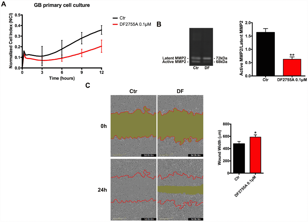
Figure 2.Cell chemotaxis assay in GB primary cell culture under DF2755A treatment. (A) Normalized cell index after 24 hours of treatment, the cell migration was followed for 12 hours. The supernatants of chemotaxis assay were collected to perform gelatin zymography. In (B) a representative gelatin zymography and relative densitometry analysis expressed as relative units of active MMP2/latent MMP2 ratio. (C) Representative images of wound closure at 0 hours (top) and 24 hours (bottom), the red lines represent the edges of the starting scratch, while the green areas represent the wound closure. The wound analysis was represented as wound width (μm) after 24 hours of migration. Data are means ± SEM of three different biological replicates (n=3). Statistical analysis was performed by the unpaired Student's t-test (with Welch’s correction). *, p< 0.05; **, p< 0.01, Ctr vs DF2755A were considered statistically significant. Ctr: Control, DF: DF2755A 0.1 μM. Scale bar = 400 μm.