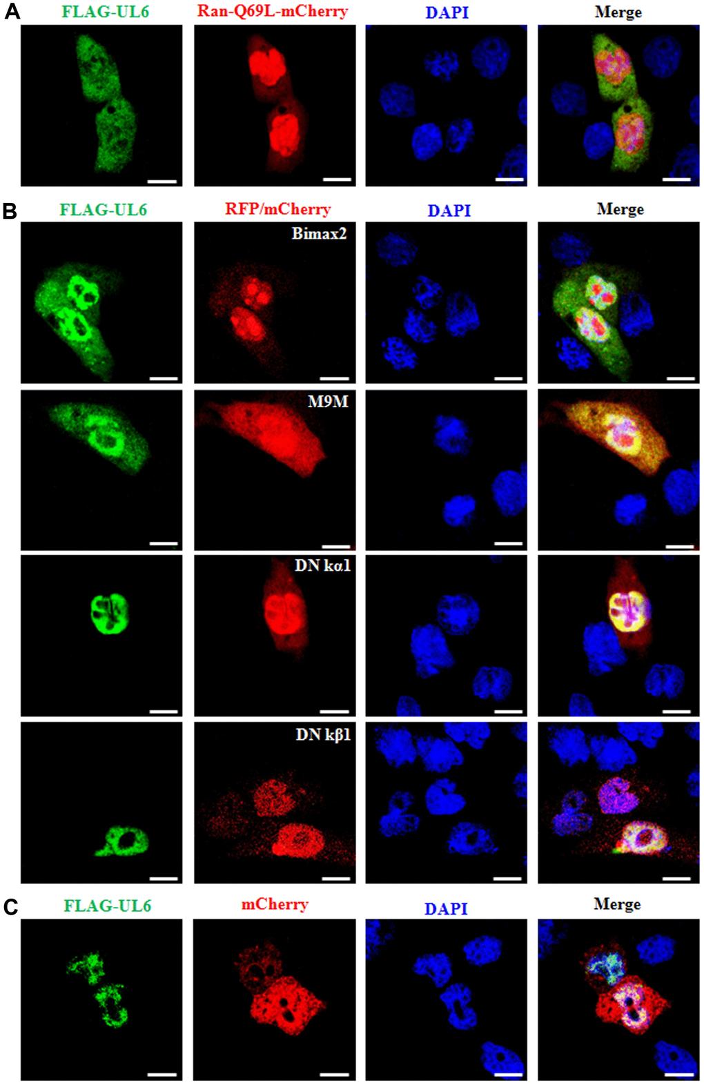
Figure 3.Nuclear import mechanism of UL6. (A) Fluorescence microscopy of COS-7 cells co-transfected with plasmids pFLAG-UL6 and pRan-Q69L-mCherry. (B) Fluorescence microscopy of COS-7 cells co-transfected with plasmid pFLAG-UL6 and plasmid encoding Bimax2-RFP, M9M-RFP, DN kα1-mCherry or DN kβ1-mCherry. (C) Fluorescence microscopy of COS-7 cells co-transfected with pFLAG-UL6 and pmCherry-N1. FITC-labeled proteins and mCherry fusion proteins were shown in its original color green and red, respectively, and the merged image was presented in yellow signal. All scale bars indicate 10 um, Statistical analysis of the fluorescence was shown in Table 3.