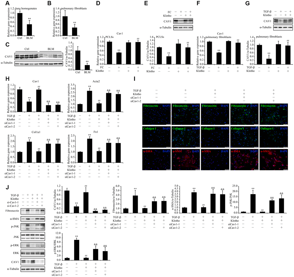
Figure 7.The mitigation of TGF-β-induced pulmonary fibroblasts activation and ECM production by Kl is partially suspended after Cav1 being silenced. mRNA levels of Cav1 were measured by qPCR in the total lung lysates (A) and isolated pulmonary fibroblasts (B) from mice 21 days after intratracheally administering a single dose of PBS (Ctrl) or bleomycin (BLM). Protein levels of CAV1 (C) were examined by western blotting in the total lung lysates from mice 21 days administering a single dose of PBS (Ctrl) or bleomycin (BLM). **P < 0.01 vs. Ctrl. 5-7 animals per group. Mouse PCLSs pre-incubated with or without mouse rKL for 24 h were randomized to be treated with control cocktail (CC) or fibrosis cocktail (FC) with or without rKL for another 48 h, when mRNA (D) and protein (E) levels of Cav1 in the mouse PCLSs from each group were measured by qPCR and western blotting, respectively. **P < 0.01 vs. CC without KL. ##P < 0.01 vs. FC without rKL. Primary pulmonary fibroblasts isolated from wild type C57BL/6 mice were pre-incubated with or without mouse rKL. After 12 h, they were randomized to be incubated with or without TGF-β and rKL for another 24 h when mRNA (F) and protein (G) levels of Cav1 were examined by qPCR and western blotting, respectively. **P < 0.01 vs. without TGF-β or KL. ##P < 0.01 vs. with TGF-β and without rKL. Primary pulmonary fibroblasts isolated from wild type C57BL/6 mice were transfected with control siRNA (siNC) or Cav1 siRNAs (siCav1-1 and -2). After siRNA transfection for 24 h, fibroblasts were pre-incubated with or without mouse rKL for 12 h, followed by treatment with or without TGF-β and rKL for another 24 h, when mRNA levels of Cav1, Acta2, Fn1, and Col1a1 were assessed by qPCR (H), fibronectin, α-SMA, and collagen I were stained by immunofluorescence staining (I, Scale bars = 100 μm), and protein levels of fibronectin, α-SMA, p-JNK, JNK, p-ERK, ERK, CAV1 and α-Tubulin were examined by western blotting (J). **P < 0.01 vs. siNC without TGF-β or rKL. ##P < 0.01 vs. siNC with TGF-β and without rKL. &&P < 0.01 vs. siNC with TGF-β and rKL.