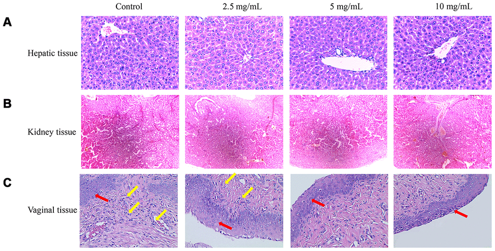
Figure 6.Therapeutic effects of flavonoids in A. conyzoides on bacterial vaginitis in mice. (A, B) HE staining showed that various doses of flavonoids in A. conyzoides did not induce obvious histomorphologic changes in liver and kidney tissues of mice. (C) In the control group, the squamous cells in the basal layer of vagina tissue were damaged and a large number of inflammatory cells were infiltrated in vagina tissue, indicating that the bacterial vaginitis model in mice was successfully established. After treatment with low concentration of flavonoids in A. conyzoides (2.5 mg/mL), fewer inflammatory cells were observed. Furthermore, after treatment with middle and high concentrations of flavonoids in A. conyzoides (5 mg/mL and 10 mg/mL), no obvious inflammatory cell infiltration in vagina tissue was observed and the squamous cells in the basal layer were arranged regularly and smoothly, indicating that inflammation in vagina tissue was remarkably improved. Red arrows point to squamous cells and yellow arrows point to inflammatory cells.