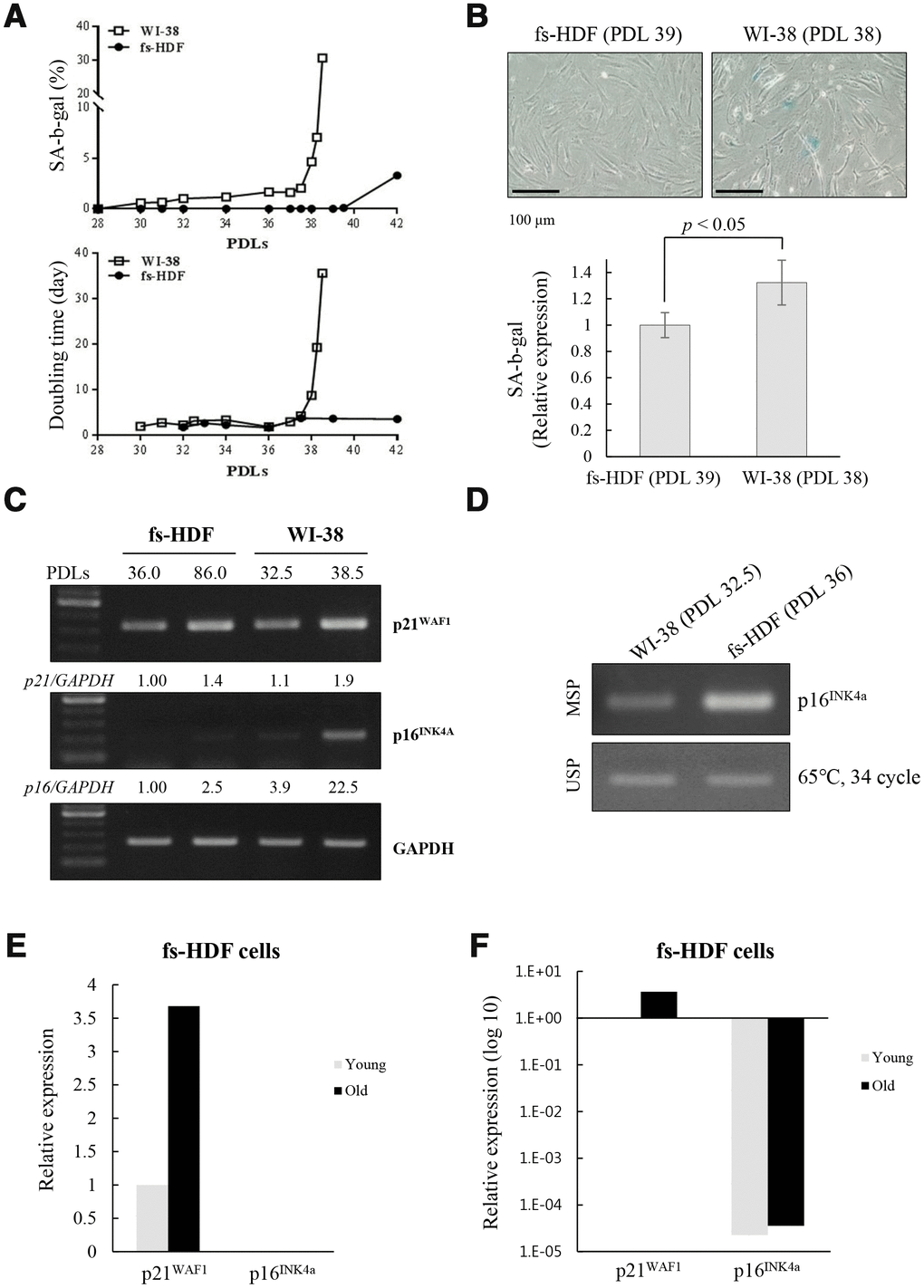
Figure 6.Replicative senescence in fs-HDF cells with p16INK4a silenced is delayed relative to WI-38 cells expressing elevated p16INK4a. (A) Earlier induction of SA-β-galactosidase in WI-38 cells compared to fs-HDF cells accompanied by significant differences in their doubling times and number of population doublings (PDLs). (B) WI-38 cells are positive for SA-β-galactosidase at 38 PDL, but fs-HDF cells at 39 PDL are not. (C) RT-qPCR analysis showing no induction of p16INK4a expression in fs-HDF cells at 86 PDL in contrast to marked induction in WI-38 cells at 38.5 PDL. (D) Methylation-specific PCR (MSP) and unmethylation-specific PCR (USP) analysis. The methylation status of the p16INK4a gene differed slightly between WI-38 and fs-HDF cells. (E) Relative expression of p21WAF1 and p16INK4a genes in old and young fs-HDF cells, as seen in RNA sequencing analysis. p16INK4a expression was almost absent in fs-HDF cells. (F) The data presented in (E) was log10 transformed. Note the absence of p16INK4a expression in young and old fs-HDF cells, in contrast to clear induction of p21WAF1 expression in the old cells.