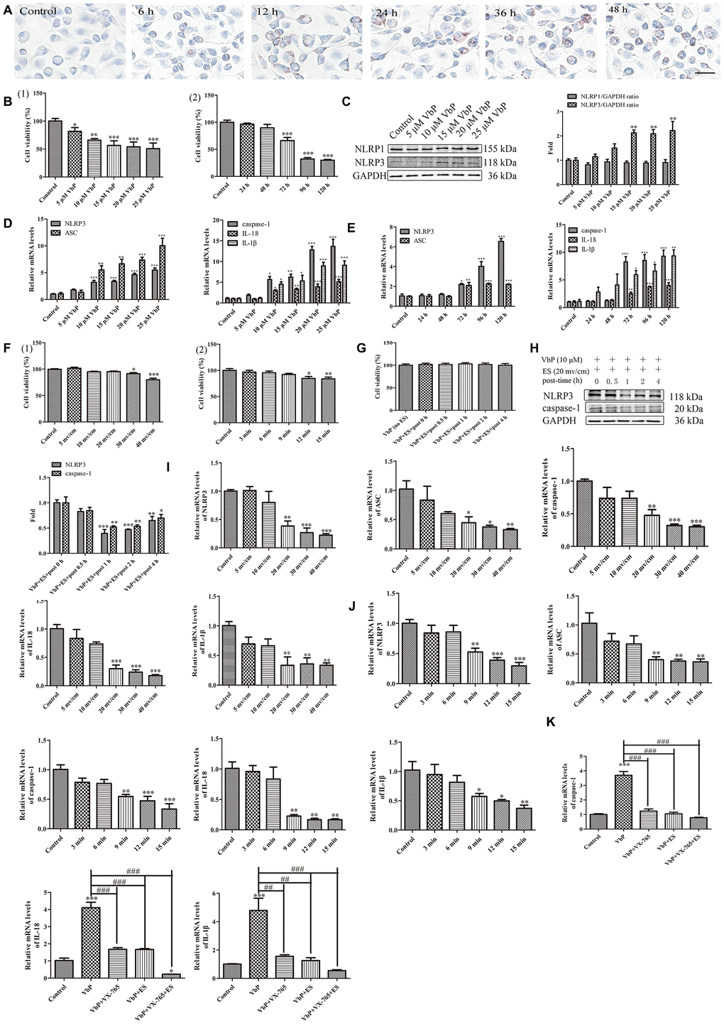
Figure 8.ES inhibits VbP-induced pyroptosis in foam cells. (A) Representative images of Oil Red O-stained THP-1 macrophages following incubation with ox-LDL for 6, 12, 24, and 48 h (scale bar: 50 μm). (B) Detection of foam cell viability by CCK8 assays: (1) Dose-response assay results (72 h incubation); (2) Time-response assay results (5 μM VbP). (C) Detection of NLRP1 and NLRP3 expression by western blotting in foam cells treated with different concentrations of VbP (72 h exposure). (D) Dose-response and (E) time-response RT-qPCR analysis of pyroptosis-related mRNAs in foam cells treated with different concentrations of VbP (72 h) or 10 μM VbP for up to 120 h. (F) Viability measurements in VbP-treated foam cells exposed to ES at (1) different voltages (9 min exposure) and (2) 20 mv/cm for various exposure times. (G) Viability measurements at different time points after ES (20 mv/cm for 9 min) in foam cells treated with VbP (10 μM for 72 h). (H) Analysis of NLRP3 and caspase-1 expression by western blotting at different time points after 9 min, 20 mv/cm ES. (I) Dose-response and (J) time-response RT-qPCR analysis of pyroptosis-related mRNAs in foam cells exposed to ES. (K) RT-qPCR analysis showing the effects of VbP, VX-765, and ES on caspase-1, IL-18, and IL-1β mRNA levels in foam cells. n = 3; *P<0.05, **P<0.01, and ***P<0.001 vs. control cells; ##P<0.01 and ###P<0.001 vs. VbP-treated cells.