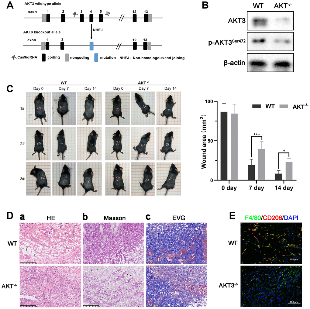
Figure 6.Loss of AKT3 delayed cutaneous wound healing in mice. (A) Schematic of AKT3 knockout in mice. (B) Western blotting for AKT3 levels in AKT3+/+ and AKT3-/- mice (n = 6). (C) AKT3 knockout delayed cutaneous wound healing in mice by days 7 and 14 post-injury. (Da–c) Histological staining of mouse cutaneous wound tissue. (a) H&E staining showed more inflammatory cells in the wound tissue of AKT3-/- mice and incomplete tissue integrity (n = 6). (b) Masson staining showed the numbers of collagenous and muscular fibers were reduced in the wound tissue of AKT3-/- mice (n = 6). (c) EVG staining showed that number of elastin fibers were decreased in the wound tissue of AKT3-/- mice (n = 6). (E) IF staining showed the F40/80 and CD206 expression in mouse cutaneous wound tissue. All the experiments were repeated at least three times.