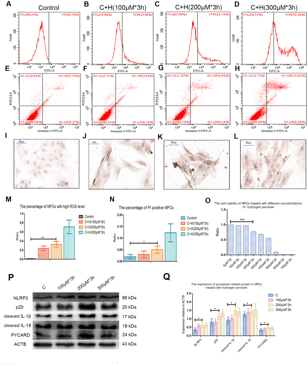
Figure 2.Hydrogen peroxide induced the pyroptosis of NPCs. (A–D) The reactive oxygen species level of the nucleus pulposus cells treated with hydrogen peroxide of 0μM, 100μM, 200μM and 300μM for 3h was detected by flow cytometry. (E–H) The corresponding apoptosis rates of nucleus pulposus cells treated with different concentrations of hydrogen peroxide were detected by flow cytometry using annexin V/PI double staining. (I–L) The immunohistochemical staining revealed the expression of CASP1 in the nucleus pulposus cells treated with different concentrations of hydrogen peroxide (magnification: ×40, scale bar = 50μm). (M) The panel showed the comparison of percentage of nucleus pulposus cells with high reactive oxygen species level after treatment with hydrogen peroxide of different concentrations. (N) The panel showed the percentage of PI positive cells measured after treatment with hydrogen peroxide with different concentrations. (O) The CCK-8 test showed the viability of the nucleus pulposus cells treated with different concentration of hydrogen peroxide. (P) The expression of NLRP3, cleaved CASP1 (p20), cleaved IL-1β, cleaved IL-18 and PYCARD in the cultured nucleus pulposus cells treated with different concentrations of hydrogen peroxide. (Q) The panel showed the averaged data measured from the images as shown in the Figure P. The data were presented as the mean ± SEM. *P < 0.05, **P < 0.01, ***P < 0.001.