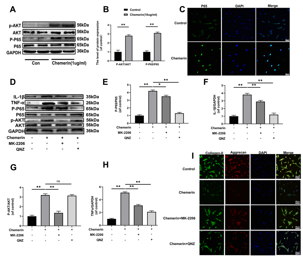
Figure 4.The AKT phosphorylation, and NF-kB signaling pathway were associated with chemerin induced cell damage. (A) The expression levels of p-AKT, AKT, p-p65, and p65 were visualized by western blotting. (B) Quantification of p-AKT, AKT, p-p65, and p65 immunoblots. (C) The expression levels of p65, and nuclear translocation were observed by immunofluorescence. (D) The expression levels of inflammatory mediators, and signaling pathway related proteins, such as IL-1β, TNF-α, p-AKT, AKT, p-p65, and p65 were analyzed by western blotting. (E–H) Quantification of IL-1β, TNF-α, p-AKT, AKT, p-p65, and p65 immunoblots. (I) Immunofluorescence of collagen II, and aggrecan in NPCs were observed by Nikon ECLIPSE Ti microscope (Nikon, Tokyo, Japan). Data are represented as mean ± SEM of three independent experiments, each done in triplicate. Significant differences between groups are indicated as **p < 0.01, *p < 0.05.