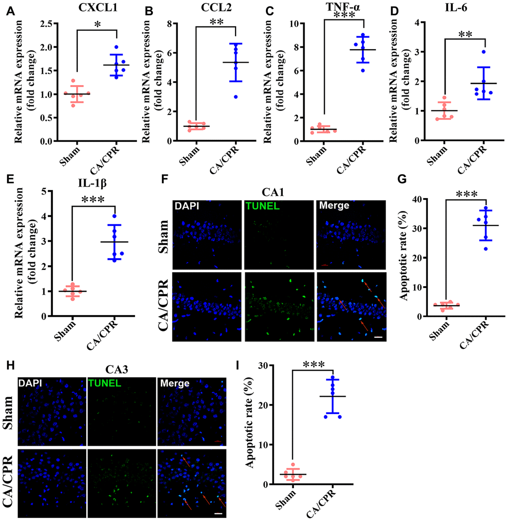
Figure 6.Neuroinflammation and neuronal apoptosis were markedly augmented in the hippocampus following CA/CPR. Hippocampus was collected at 4h and 72h after resuscitation for the detection of neuronal apoptosis and inflammatory cytokines, respectively. (A–E). The levels of CXCL1 (A), CCL2 (B), TNF-α (C), IL-6 (D), and IL-1β mRNA (E). (F). Representative images for TUNEL detection in the hippocampal CA1 subarea. (G). The percentage of TUNEL-positive cells in the CA1 region. (H). Representative images for TUNEL detection in the hippocampal CA3 subarea. (I). The percentage of TUNEL-positive cells in the CA3 region. The cells pointed by the red arrows represent typical TUNEL-positive cells. n = 6 per group. **P < 0.01, ***P < 0.001. CXCL1, chemokine (C-X-C motif) ligand 1; CCL2, chemokine (C-C motif) ligand 2; TNF-α, tumor necrosis factor α; IL-1β, interleukin-1β; IL-6, interleukin-6.