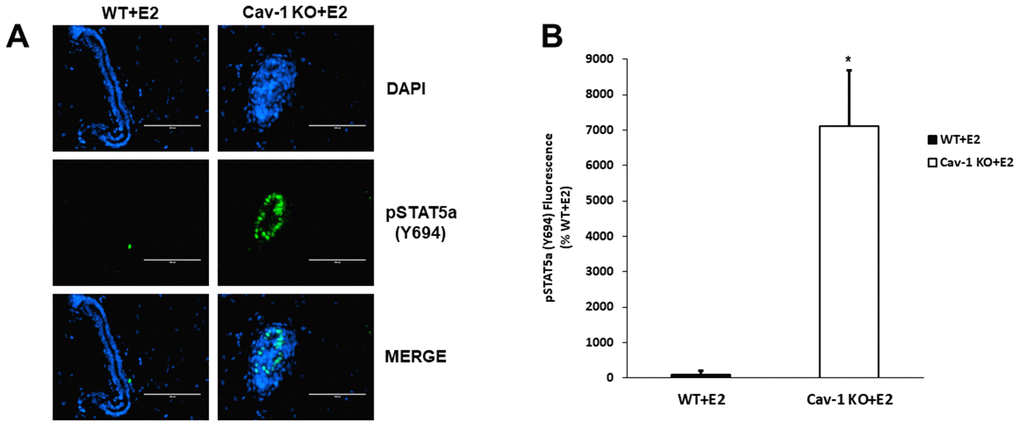
Figure 1.Cav-1 KO DCIS lesions display increased phosphorylated STAT5a (Y694) levels as a response to 17β-Estradiol treatment. (A) Mammary glands of estrogen-treated WT and Cav-1 KO mice were immunostained with an antibody recognizing phosphorylated STAT5a (Y694). DAPI was used as a nuclear counterstain. The EVOS FL microscope was used to capture images at 40x objective with the DAPI and CY5.5 light cubes (blue: DAPI immunostaining; green: phosphorylated STAT5a (Y694) immunostaining). For each experimental group, immunofluorescence was performed in triplicate on mammary glands derived from 3 independent mice. (B) Immunofluorescence staining was quantified using Image J software. Corrected total cell fluorescence (CTCF) was calculated using the following formula: CTCF = Integrated Density – (Area of Selected Region x Mean Fluorescence of Background). Cav-1 KO mammary glands demonstrated a significant increase in phosphorylated STAT5a (Y694) expression compared to estrogen-treated WT counterparts (71.1-fold, p<0.05, n=3). Data are expressed as % WT+E2.