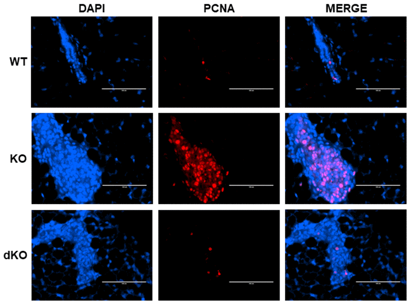
Figure 3.Proliferating Cell Nuclear Antigen (PCNA) increase in Cav-1 KO DCIS-like lesions secondary to estrogen treatment is inhibited by a homozygous STAT5a deletion. Mammary glands of estrogen-treated WT, Cav-1 KO, and Cav-1/STAT5a dKO mice were immunostained with an antibody recognizing proliferating cell nuclear antigen (PCNA). DAPI was used as a nuclear counterstain. The EVOS FL microscope was used to capture images at 40x objective with the DAPI and Texas Red light cubes (blue: DAPI immunostaining; red: PCNA immunostaining). Qualitatively, mammary glands lacking Cav-1 expression showed elevated PCNA expression upon stimulation with estrogen compared to WT counterparts. A STAT5a deletion in the Cav-1 KO mammary gland diminished PCNA expression to WT levels. For each experimental group, immunofluorescence was performed in triplicate on mammary glands derived from 3 independent mice.