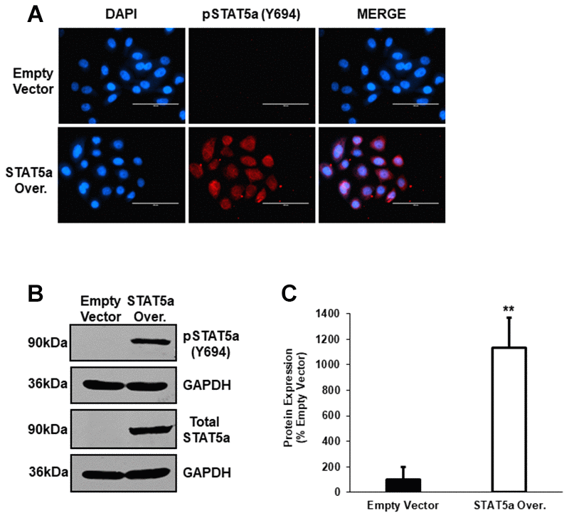
Figure 5.Western blot analysis following lentiviral-mediated overexpression of STAT5a in human MCF10DCIS.com. (A) Immunofluorescence staining was used to confirm overexpression of phosphorylated STAT5a (Y694) in MCF10DCIS.com cells. Empty vector and STAT5a overexpressor cells were immunostained with an antibody recognizing phosphorylated STAT5a (Y694). DAPI was used as a nuclear counterstain. The EVOS FL microscope was used to capture images at 40x objective with the DAPI and Texas Red light cubes (blue: DAPI immunostaining; red: phosphorylated STAT5a (Y694) immunostaining). Qualitatively, phosphorylated STAT5a (Y694) expression was upregulated in STAT5a overexpressor cells compared to empty vector control cells. Immunofluorescence was performed in triplicate on cells derived from 3 independent passages. (B) Western blotting was used to confirm overexpression of phosphorylated STAT5a (Y694) in MCF10DCIS.com cells. Whole cell lysates (100μg) of empty vector and STAT5a overexpressor cells were used to assess the protein expression of phosphorylated STAT5a (Y694) and total STAT5a. GAPDH was used as a control for equal loading. Western blotting was performed in triplicate on cells derived from 3 independent passages. (C) Densitometry analysis was performed using the LI-COR imager. A ratio of phosphorylated STAT5a (Y694) to total STAT5a was calculated upon normalizing to respective loading controls. Data are reported as % empty vector. Quantitatively, phosphorylated STAT5a (Y694) expression was upregulated in MCF10DCIS.com STAT5a overexpressor cells compared to empty vector control cells (11.3-fold, p<0.01, n=3).
Figure 5 — Essential role of STAT5a in DCIS formation and invasion following estrogen treatment | Aging