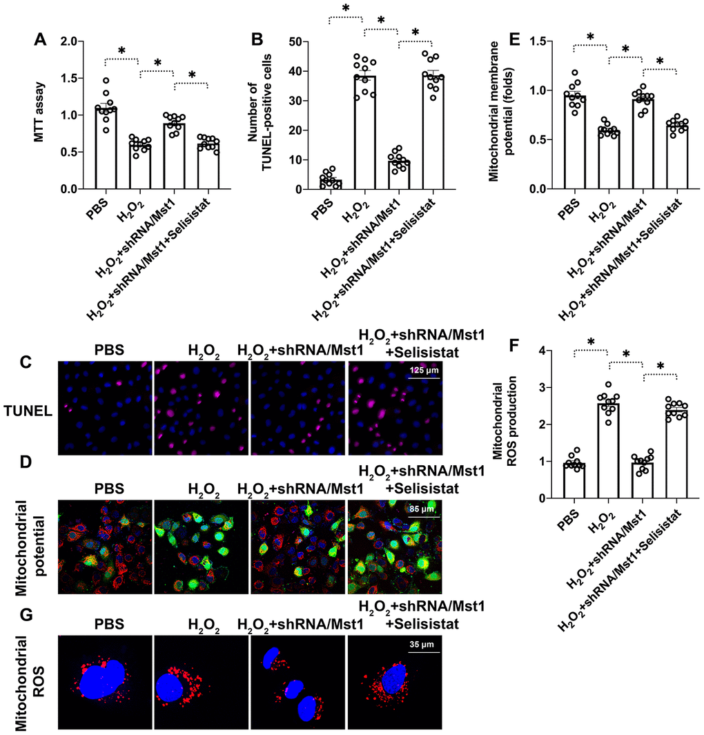
Figure 4.Sirt1 inhibition abolishes the beneficial effects of Mst1 knockdown in oxidative stress-induced RAFLSs. (A) MTT assay results show the viability of control and Mst1-knockdown RA-FLSs, pretreated with or without selisistat, a potent inhibitor of Sirt1. (B, C) TUNEL assay results show the apoptotic rates (percent Tunel-positive cells) in the control and Mst1-knockdown RA-FLSs, pretreated with or without selisistat, and treated with or without 0.3 mM H2O2 for 6 h. (D, E) JC-1 staining assay results show mitochondrial membrane in the control and Mst1-knockdown RA-FLSs, pretreated with or without selisistat, and treated with or without 0.3 mM H2O2 for 6 h. Mitochondrial potential was measured by the ratio of red-to-green JC-1 fluorescence intensity. (F–G) Representative fluorescence microscopic images show the DCFDA staining to determine ROS levels in the control and Mst1-knockdown RA-FLSs, pretreated with or without selisistat, and treated with or without 0.3 mM H2O2 for 6 h. ROS levels were quantified based on DCFDA staining intensity. *P<0.05.