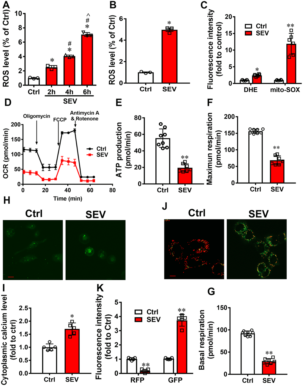
Figure 1.Sevoflurane induced mitochondria impairment in vitro. The ROS levels of H4 cells were tested after treatment with 4.1% sevoflurane for 2 h, 4 h, and 6 h (SEV-2h, SEV-4h, and SEV-6h, respectively) (A) H4 cells were treated with 4.1% sevoflurane for 6 h and ROS level was measured after 24 h. (B) The intracellular ROS and mitochondrial ROS levels were measured after treatment with 4.1% sevoflurane for 6 h (C) The OCR assay was used to observe the mitochondrial respiratory function in H4 cells after treatment with 4.1% sevoflurane for 6 h (SEV) (D–G). The cytoplasmic calcium levels were detected using a Fluo-4 AM probe (H, I) and the membrane potential was measured using a JC-1 probe (J, K). Images show representative examples from three independent experiments for each group. The data are expressed as mean ± SD. (A) *P<0.05, Ctrl vs SEV-2h, SEV-4h and SEV-6h group; # P<0.05, SEV-2h vs SEV-4h and SEV-6h group; ^ P<0.05, SEV-4h vs SEV-6h group. (B) * P<0.05, Ctrl vs SEV-6h group. (C, E, F, G, I, K) *P<0.05, **P<0.05, Ctrl vs SEV group.