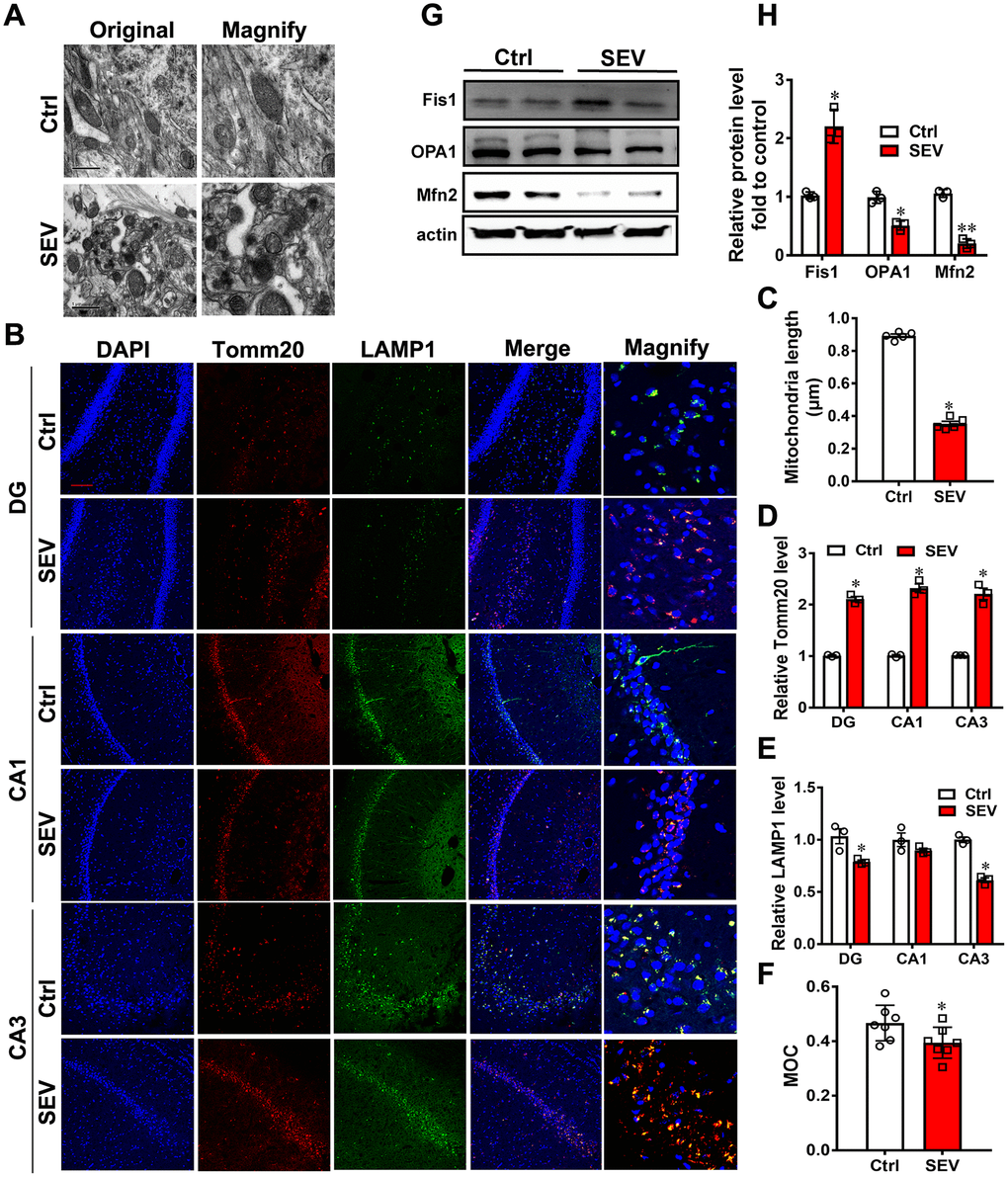
Figure 2.Sevoflurane induced mitochondria impairment in vivo. Eighteen-month-old rats were subjected to 2% sevoflurane for 5 h (SEV). After perfusion, the ultrastructure of mitochondria in the hippocampus was observed under an electron microscope. (A) The lengths of all mitochondria were measured and are shown in (C) Tomm20 and LAMP1 protein levels in the hippocampus were measured by immunofluorescence assay. Scale bar represents 50 μm. (B) The levels of Tomm20 are shown in (D) and LAMP1 in (E) Manders’ overlap coefficient (MOC) was calculated to determine the degree of colocalization. (F) The proteins Fis1, OPA1 and Mfn2 were examined by western blotting. (G) The results of semi-quantitative analysis of Fis1, OPA1, Mfn2 and β-actin are shown in (H) Images show representative examples from three independent experiments for each group. The data are expressed as mean ± SD. (C-R) * P<0.05, **P<0.01, Ctrl vs SEV.