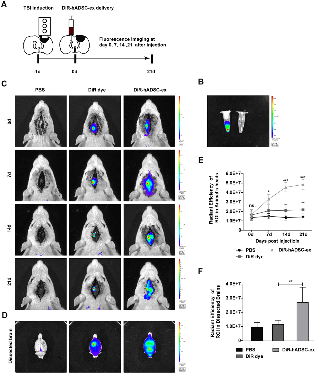
Figure 3.Visualization and in vivo tracking of hADSC-ex after intracerebroventricular administration. (A) Schematic representation of the experimental procedures. (B) Representative fluorescence images of DiR-labeled hADSC-ex and PBS. (C) Representative fluorescence images of rat heads on days 0, 7, 14 and 21 after administration of PBS, DiR dye or DiR-hADSC-ex. (D) Representative fluorescence images of dissected brains on day 21. (E) Fluorescence intensity quantification of regions of interest in the lesion sites of rat heads, expressed as the average radiance ± SD, n = 5 rats per group. ns. p > 0.05, * p < 0.05, *** p < 0.001, determined by repeat-measures two-way ANOVA vs. DiR dye control group. (F) Fluorescence intensity quantification of regions of interest in the lesion sites of dissected rat brains, expressed as the average radiance ± SD, n = 5 rats per group. ns. p > 0.05, ** p < 0.01, determined by one-way ANOVA vs. DiR dye control group.