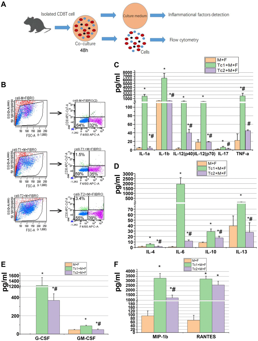
Figure 4.Tc1-treated macrophages promoted the activation of fibroblast to secrete inflammatory factors. (A) CD8+ T cells (Tc1 and Tc2) were isolated from UUO kidneys and cocultured with Raw264.7 cells plus NIH3T3 cells for 48 h (1 × 10⁴ T cells, 1 × 10⁵ Raw264.7 cells, and 2 × 10⁵ NIH3T3 cells per well). Cell culture medium was collected for inflammatory factor detection, and cells were collected for flow cytometry. (B) Representative examples of the FACS analysis of cocultured cells. Cells were stained with CD8 and F4/80 and sorted through flow cytometry; then, NIH3T3 cells (CD8−F4/80− cells) were collected for the examination of α-SMA, Col-1, and fibronectin mRNA (data shown in Figure 5). (C–F) Inflammatory factors were evaluated by using a Luminex multiplex murine cytokine assay, and those that were significantly elevated are shown (*p < 0.05 vs. Raw264.7 cells + NIH3T3 cells, #p < 0.05 vs. Tc1 + Raw264.7 cells+ NIH3T3 cells).