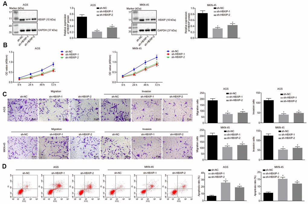
Figure 2.Silencing HBXIP inhibits GC cell viability, migration and invasion, and induces apoptosis. (A) Representative Western blots of HBXIP protein and its quantitation in AGS and MKN-45 cells upon HBXIP silencing, normalized to GAPDH. (B) CCK-8 was used to examine the proliferation of AGS and MKN-45 cells upon HBXIP silencing. (C) Transwell assay was used to examine the migration and invasion ability of AGS and MKN-45 cells upon HBXIP silencing (× 200). (D) Flow cytometry was used to examine the apoptosis of AGS and MKN-45 cells upon HBXIP silencing. * p < 0.05 vs. the sh-NC group (AGS or MKN-45 cells treated with sh-NC). The above data were measurement data, and expressed as mean ± standard deviation. Data in panels (A, C and D) were analyzed by one-way ANOVA with Tukey’s post hoc test and in panel B by repeated measures ANOVA with Bonferroni post hoc test. The cell experiment was repeated 3 times independently.