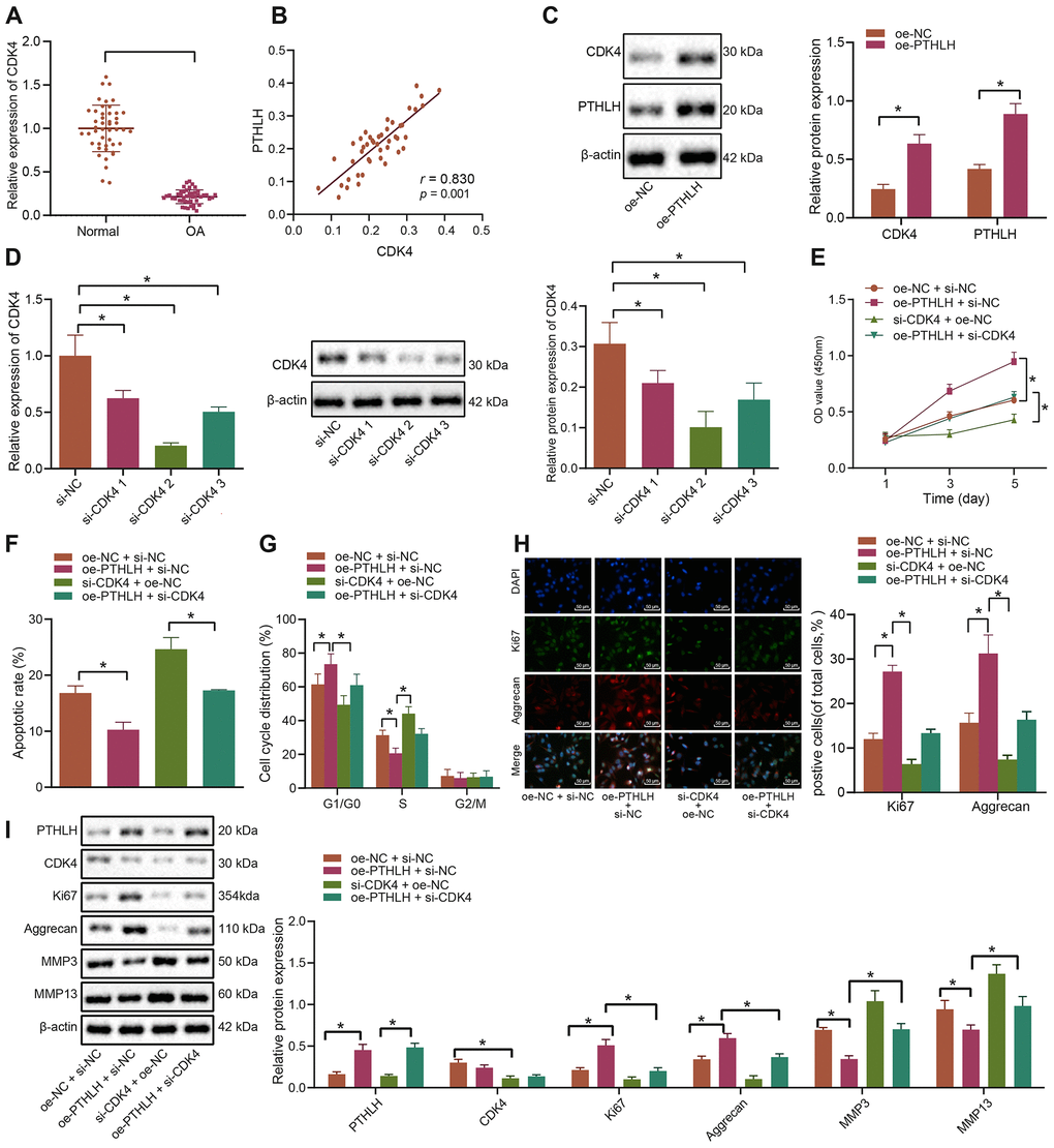
Figure 4.PTHLH decelerates the apoptosis of IL-1β-treated chondrocytes by enhancing CDK4. (A) The mRNA expression of CDK4 in OA cartilage tissues (n = 46) and normal cartilage tissues (n = 46) determined by RT-qPCR. (B) Pearson’s analysis of the correlation between PTHLH and CDK4 expression. (C) The protein expression of CDK4 in chondrocytes in response to overexpression of PTHLH normalized to β-actin measured by Western blot analysis. (D) The expression of CDK4 after siRNA transfection measured by RT-qPCR and Western blot analysis (normalized to β-actin). In panel (E–I) IL-1β-treated chondrocytes were co-transfected with oe-PTHLH/oe-NC and si-CDK4/si-NC. (E) Viability of IL-1β-treated chondrocytes evaluated by MTT assay. (F) Apoptosis of IL-1β-treated chondrocytes assessed by flow cytometry. (G) Cell cycle distribution of IL-1β-treated chondrocytes assessed by flow cytometry. (H) Immunofluorescence staining of Ki67 and Aggrecan in IL-1β-treated chondrocytes (× 200, scale bar = 50 μm). (I) The protein expression of Ki67, Aggrecan, MMP3, MMP13 in IL-1β-treated chondrocytes normalized to β-actin measured by Western blot analysis. *p < 0.05. Statistical data were measurement data and described as the mean ± standard deviation. The independent sample t-test was conducted for comparison between the two groups. The one-way ANOVA was used for comparison among multiple groups followed by Tukey’s post hoc test. The experiment was repeated 3 times independently.