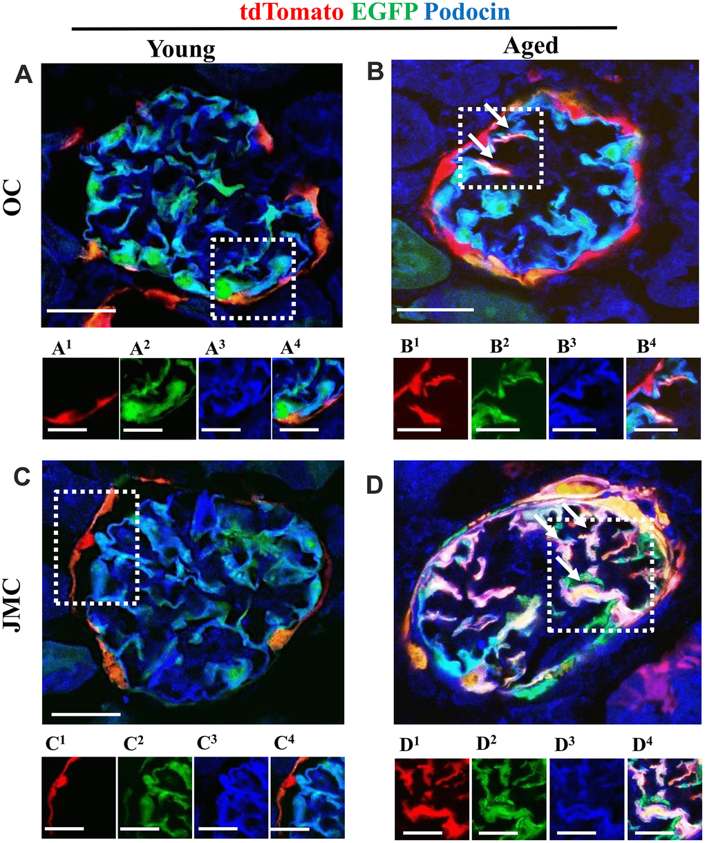
Figure 5.A subset of newly generated podocytes (tdTomato+EGFP+) from PEC origin co-express podocin in the glomerular tuft of aged mice. (A–D) Representative confocal images of tdTomato+ (red), EGFP+ (green) and podocin staining (blue) in young and aged mice. The inserts show separate channels of the outlined glomeruli, with superscripts: 1=tdTomato, 2=EGFP, 3=podocin and 4=merged. Podocin staining was detected with an antibody, tdTomato and EGFP reporters were detected without antibody. (A) Young mice (OC) showed that tdTomato+PECs (red) are detected along Bowman’s capsule (A1). The majority of EGFP+ cells (green) (A2) co-localize with podocin (blue) (A3) and create a cyan color (A4). (B) Aged mice (OC) showed that a subset of differentiated tdTomato+ PECs (red) (B1) co-expresses EGFP+ (green) (B2) and podocin (blue) (B3) creating a pink/white color in the glomerular tuft (white arrows) (B4). (C) Young mice (JMC) showed that tdTomato+PECs (red) (C1) are detected along Bowman’s capsule. The majority of EGFP+ cells (green) (C2) co-localize with podocin (blue) (C3) in the glomerular tuft creating a cyan color (C4). (D) Aged mice (JMC) showed that a higher number of tdTomato+PECs (red) (D1) have migrated to the glomerular tuft (marked with white arrows), become EGFP+ (D2) and co-localized with podocin (blue) (D3), creating a pink/white color (D4). Scale bars represent 25μm or 5μm (insets).