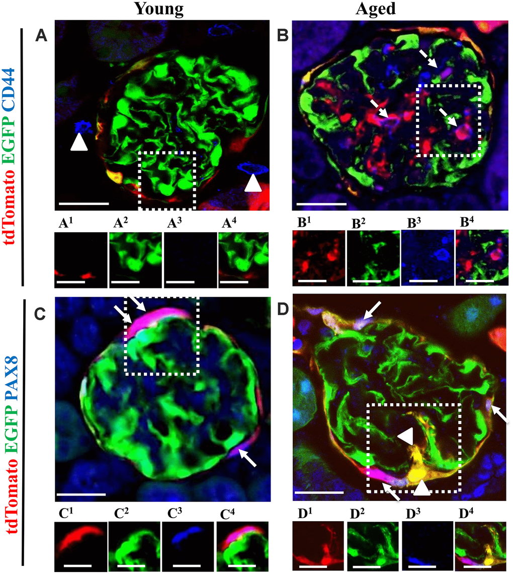
Figure 9.The majority of tdTomato+PECs co-express PAX8 along Bowman’s capsule, however newly generated podocytes from PEC origin no longer express PAX8. Activated PECs (tdTomato+CD44+) were detected in the glomerular tuft of aged mice. (A, B) Representative confocal images of tdTomato+ (red), EGFP+ (green) and CD44+ (blue) in young and aged mice. The inserts show separate channels of the outlined glomeruli, with superscripts: 1=tdTomato, 2=EGFP, 3=CD44 and 4=merged. (A) Young mice showed that CD44 staining was not detected in the glomerular tuft. An occasional CD44+ cell was observed around glomeruli (marked with arrow heads). Each channel for the glomerular tuft region marked with the dashed box are presented in panels (A1–A4). (B) Aged mice showed that a subpopulation of tdTomato+PECs (B1) that migrated to glomerular tuft were positive for CD44 (magenta color, B3- B4), marked with dashed arrows, with no overlap with EGFP (B2). (C, D) Representative confocal images of tdTomato+ (red), EGFP+ (green) and PAX8 (blue) in young and aged mice. The inserts show separate channels of the outlined glomeruli, with superscripts: 1=tdTomato, 2=EGFP, 3=PAX8 and 4=merged. (C) Young mice showed that PAX8 staining (C3) was detected in PECs along Bowman’s capsule and co-localized with tdTomato reporter (C1) creating a pink/purple color (C4) (marked with white arrows). PAX8 was not detected in podocytes (EGFP+ cells) (C2). (D) Aged mice showed that the majority of tdTomato+PECs (D1) co-express PAX8 (D3) creating a magenta/white color (D4) (marked with solid arrow). However, newly generated podocytes from PEC origin (tdTomato+EGFP+) (D1, D2) do not express PAX8 in the glomerular tuft of aged mice (marked with arrow heads). Scale bars represent 25μm or 5μm (insets).