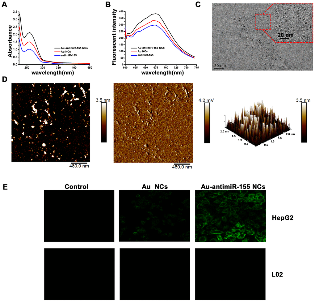
Figure 2.Characterization of in situ assembled fluorescent Au-antimiR-155 NCs. (A) UV absorption spectra of Au-antimiR-155 complexes extracted from HepG2 cells cultured with gold precursor and miR-155 inhibitors. (B) Fluorescence spectra of the aqueous solution of the extracted Au-antimiR-155 complexes. The relevant emission peak is centered at ~680 nm after excitation (EX) at 580 nm. (C) Representative TEM image of the isolated Au-antimiR-155 NCs. (D) Representative AFM image (left panel) and 3D model diagram (right panel) of biosynthetic Au-antimiR-155 NCs. The AFM phase diagram shown in the middle panel allows distinction of antimiR-155 and Au-antimiR-155 NCs. (E) Laser confocal fluorescence images of control (DMEM) cells and cells cultured in the presence of gold precursor alone (Au NCs) or gold precursor plus antimiR-155 (Au-antimiR-155 NCs). In the above experiments, the concentration of gold precursor solution was 5 μM and the concentration of antimiR-155 was 100 nM.