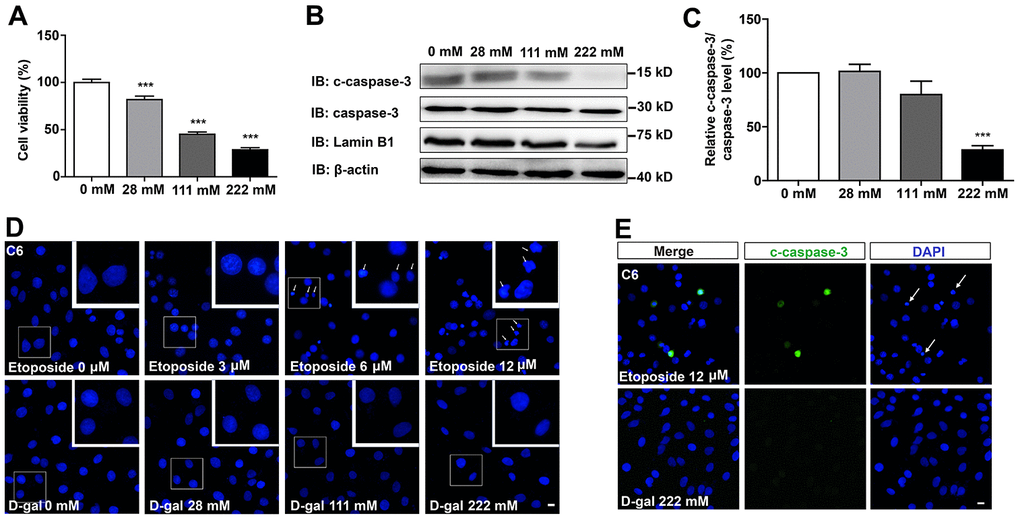
Figure 1.D-gal decreased cell viability, but did not induce apoptosis of GBM cells. (A) The effects of D-gal (treated for 8 d) on C6 cell viability as detected by CCK8 (n=3). (B) Western blot detected the expression of c-caspase-3, caspase-3 and Lamin B1 in cells treated with 0 mM, 28 mM, 111 mM and 222 mM D-gal for 8 d. (C) Quantification of c-caspase-3/caspase-3 protein level as shown in (B) (n=4). “Relative c-caspase-3/total caspase-3 level” of the 0 mM group = [c-caspase-3/total caspase-3 (0 mM group)]/[c-caspase-3/total caspase-3 (0 mM group)] ×100% = 100%; “Relative c-caspase-3/total caspase-3 level” of other D-gal-treated groups = [c-caspase-3/total caspase-3 (other D-gal-treated groups)]/[c-caspase-3/total caspase-3 (0 mM group)] ×100%. (D) DAPI staining of C6 cells treated with 0 μM, 3 μM, 6 μM, 12 μM etoposide for 1 d, and recovered for 4 d or with D-gal at 0 mM, 28 mM, 111 mM and 222 mM for 8 d. The white arrows indicated apparently dead cells. (E) Immunostaining of c-caspase-3 in cells treated with 12 μM etoposide for 1 d, and recovered for 4 d, or with D-gal at 222 mM for 8 d. The white arrows indicated apparently dead cells. Scale bars, 20 μm. Data shown are mean ± s.e.m. ***P < 0.001.