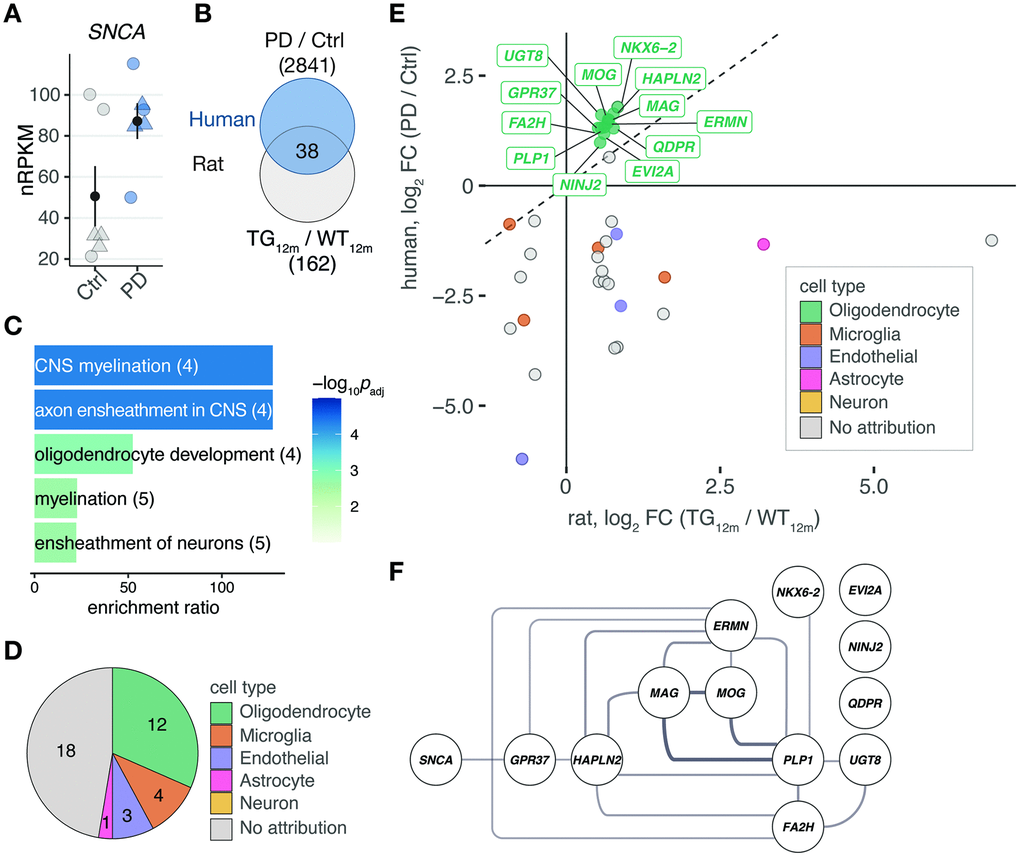
Figure 4.Shared differentially expressed genes in the rat model overexpressing SNCA and PD patients point to myelination and oligodendrocytes. (A) Expression changes of SNCA in PD patients compared to controls plotted as individual data points with mean ± SEM. Circles represent females, rectangles males. (B) Venn diagram comparing 162 DEGs identified in 12-month-old TG rats and 2841 DEGs identified in frontal cortex of PD patients according to cut-offs of padj ≤ 0.1 and │log2FC│≥ 0.5. (C) Overrepresented biological processes among 38 DEGs shared between rat and human (see B). Five most significant terms, their adjusted p-values, enrichment ratios, and underlying gene count shown. (D) Pie chart showing attribution of 38 DEGs shared between rat and human to cell types according to reference data from McKenzie et al. [19]. (E) Scatter plot of 38 DEGs identified in frontal cortex of rat and human. Cell type attributions color-coded. Oligodendrocyte DEGs labelled. (F) Protein-protein interaction network derived from 38 DEGs attributed to oligodendrocytes plus SNCA. Interactions according to String database. Only connected nodes shown. Line width reflects String interaction score.