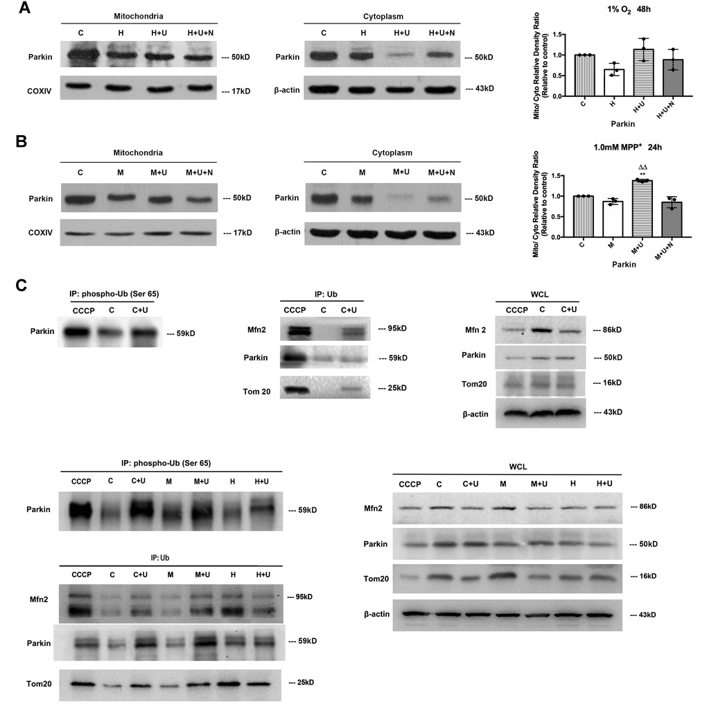
Figure 5.DOR activation promoted Parkin’s translocation from cytoplasm to mitochondria and its phosphorylation at Ser65 UBL domain and increased OMM ubiquitination for mitophagy. (A) PC12 cells were exposed to hypoxia at 1% O2 for 48 hrs, the protein extracted from mitochondria and cytosols were analyzed by Western blot respectively. C: normoxic control. H: hypoxia. H+U: DOR was activated using UFP-512 in hypoxic conditions. H+ U+N: PC12 cells were treated with UFP-512 plus naltrindole at the same time in hypoxic conditions. N=3 for each group. Note that hypoxia at 1% O2 for 48 hrs led to a significant decrease in Parkin expression both in the mitochondria and cytosol. Activating DOR using UFP-512 caused a modest increase in the ratio of mitochondria/plasma Parkin density, appearing as a sharp decrease of Parkin in the cytoplasm and an inappreciable increase of Parkin in the mitochondria. (B) PC12 cells were exposed to 1.0mM MPP+ for 24 hrs. The Parkin expressed both in mitochondria and cytosols were measured using Western blot. C: control. M: MPP+. M+U: DOR was activated using UFP-512 and exposed to MPP+. M+U+N: PC12 cells were treated with UFP-512 plus naltrindole and exposed to MPP+. N=3 in each group. **p<0.01 vs. control. ΔΔp<0.01 vs. M. Note that DOR activation caused a translocation of Parkin from cytoplasm to mitochondria with a significant decrease of Parkin in cytosol and a noticeable increase of Parkin in mitochondria under MPP+ insults. (C) The PC12 cells were treated with CCCP or exposed to hypoxia at 1% O2 for 48 hrs or 1.0 mM MPP+ for 24 hrs and the control group were established. The proteins were immunoprecipitated with anti-Ub antibody or anti-phospho-Ub (Ser65) antibody. Immunoprecipitants (IPs) and whole cell lysates (WCLs) were analyzed for Parkin, Mfn2 and Tom20. CCCP: positive control. PC12 cells were treated with 10 μM CCCP for 24 hrs. C: control. C+U: the cells were treated with UFP-512. H: hypoxia. H+U: DOR activation with UFP-512 in hypoxic condition. M: MPP+. M+ U: DOR was activated using UFP-512 and then exposed to MPP+. Note that the administration of UFP-512 promoted the phosphorylation of Parkin at its Ser65 UBL domain, and increased the ubiquitination of Mfn2 and Tom20 both under normal conditions and MPP+ insults. Hypoxia induced a remarkable degradation in Mfn2 and Tom20 expression with an increase in the ubiquitination of these two proteins. UFP-512 did not appreciably alter the hypoxia-mediated effects.