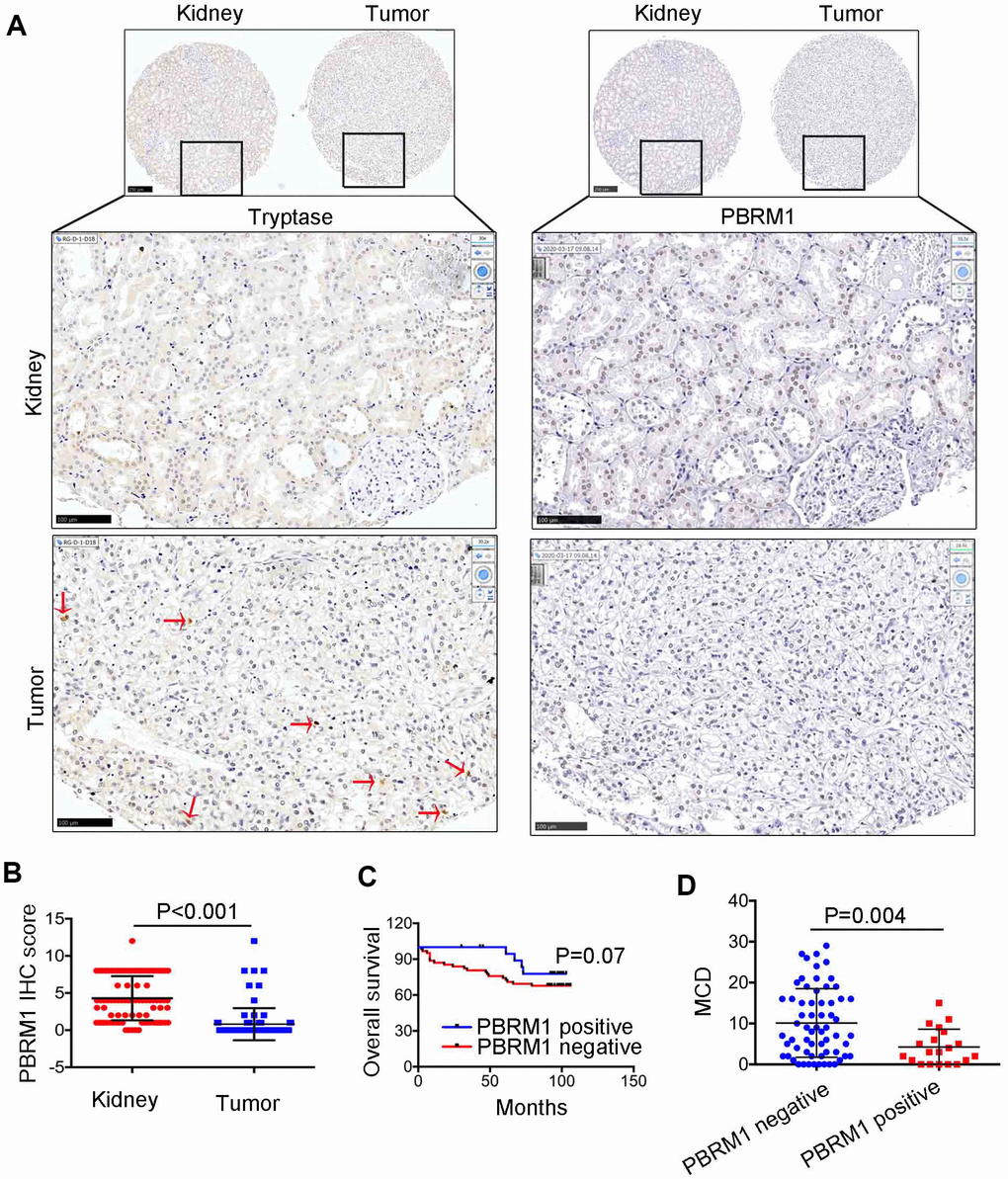
Figure 4.The relationship between PBRM1 protein expression and mast cell infiltration in ccRCC based on IHC analysis. (A) Representative immunohistochemical images show PBRM1- and tryptase-positive mast cells in ccRCC and adjacent normal kidney tissue samples. (B) Dot plot of PBRM1 IHC staining score in adjacent normal kidney tissues (n=83) and ccRCC tissues (n=83). (C) Overall survival of ccRCC patients with PBRM1 IHC staining negative group (n=65) or PBRM1 IHC staining positive group (n=20). (D) Pearson correlation analysis shows the association between PBRM1 expression and mast cell infiltration in 85 out of 90 ccRCC patient tumor tissue samples. Data for five tumor tissues is not included (missing the tissues in TMA). Note: Statistical significance was based on Student’s t-test.