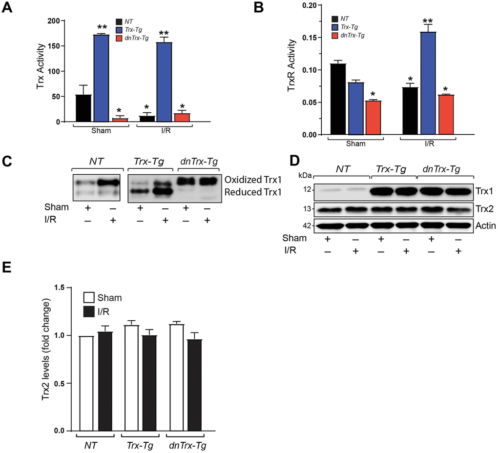
Figure 1.High amounts of hTrx in transgenic mice prevents I/R mediated redox shift, and loss of Trx and Trx reductase activities. (A) Trx activity was assayed in myocardium derived from sham and I/R-subjected NT, Trx-Tg, and dnTrx-Tg mice and expressed as nanomoles of NADPH oxidized per minute per milligram of protein at 25°C. Values are represented as means ± SEM (n =3-4). *p <0.05 versus NT sham; **p <0.05 versus NT or dnTrx-Tg. (B) TrxR activity in sham or I/R myocardium were expressed as micromoles of 5-thio-2-nitrobenzene (TNB) formed per minute per milligram of protein at 30°C. Values are represented as means ± SEM (n =3-4). *p <0.05 versus NT sham; **p <0.05 versus NT or dnTrx-Tg I/R. (C) Redox Western blot analysis revealing the redox state of Trx (oxidized and reduced) in sham or I/R myocardium from NT, Trx-Tg and dnTrx-Tg mice. (D) AAR region of sham or I/R myocardium from NT, Trx-Tg and dnTrx-Tg were lysed using M-PER lysis buffer and analyzed for Trx1, Trx2 and Actin by western blotting. (E) Trx2 levels were quantified and expressed as fold change. Statistical significance was determined with one-way ANOVA followed by Tukey’s post-hoc multiple comparisons test.