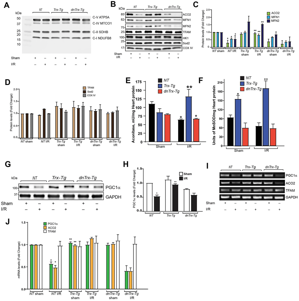
Figure 6.Trx prevents I/R-induced loss of mitochondrial proteins, by upregulating transcription of PGC1α. (A). Mitochondria was isolated from sham or I/R subjected NT, Trx-Tg and dnTrx-Tg mice. The mitochondrial extracts were analyzed for oxidative phosphorylation complex subunits by western blot using Abcam OXPHOS cocktail antibody. (B) Western blot analysis of ACO2, MFN1, MFN2, TFAM, Hexokinase 1 (HK-1), Sod2 and COX IV in sham or I/R mitochondrial extracts from NT, Trx-Tg and dnTrx-Tg mice. (C, D). Protein levels were quantified and expressed as fold change. *p <0.05 versus NT sham; **p <0.05 versus NT or dnTrx-Tg I/R. (E) Aconitase 2 activity and (F). MnSOD activity was determined in sham and I/R myocardium obtained from NT, Trx-Tg, or dnTrx-Tg mice as described in materials and methods. *p <0.05 versus respective sham; **p <0.05 versus NT or dnTrx-Tg I/R. n=3. (G) AAR region of sham or I/R myocardium from NT, Trx-Tg and dnTrx-Tg were lysed using M-PER lysis buffer and analyzed for PGC1α and GAPDH by western blotting. (H). Level of PGC1α was quantified and expressed as fold change. *p <0.05 versus NT sham; **p <0.05 versus NT or dnTrx-Tg I/R. (I). RT-PCR analysis of PGC1α, ACO2 and TFAM in sham and I/R myocardium (J). mRNA levels of PGC1a, ACO2 and TFAM were quantified and expressed as fold change. *p <0.05 versus NT sham; **p <0.05 versus NT or dnTrx-Tg I/R. Statistical significance was determined with the Student’s t test (C, D, H, and J) and one-way ANOVA followed by Tukey’s post-hoc multiple comparisons test (E, and F).