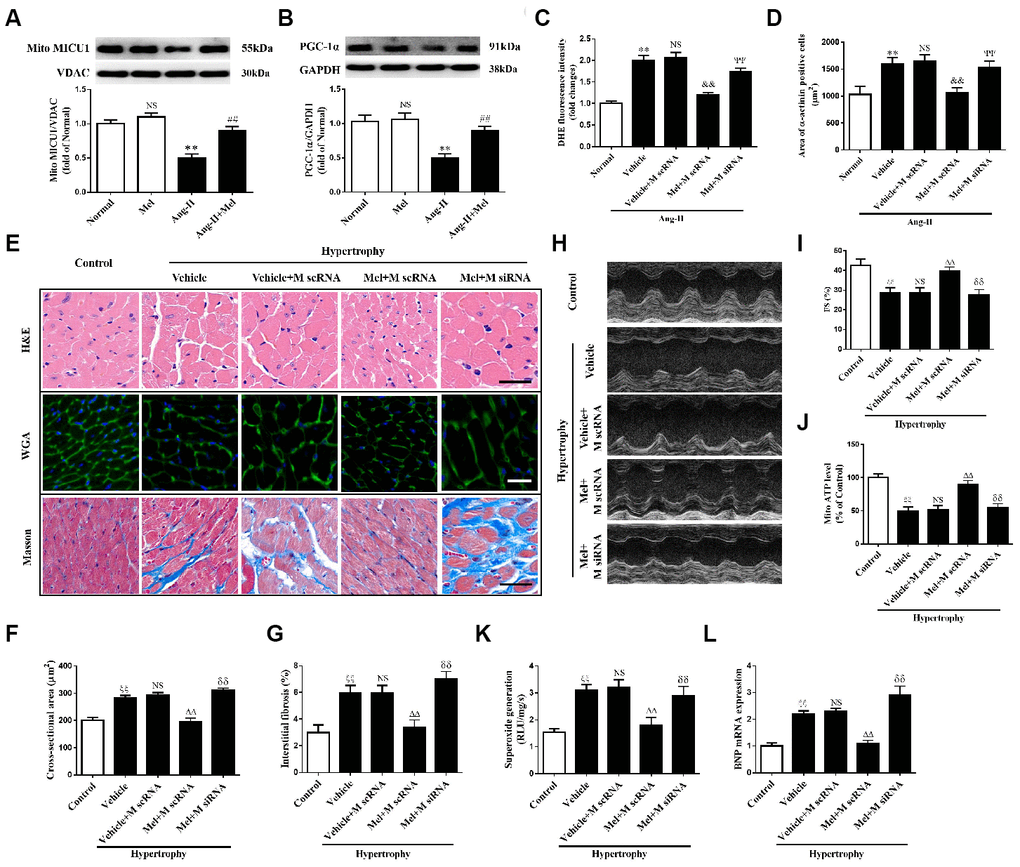
Figure 8.Melatonin ameliorated Ang-II-induced cardiac hypertrophy by increasing MICU1 pathway. (A, B) Western blotting was used to determine expression levels of MICU1 (A) and PGC-1α (B) in NMVMs with the treatment of Ang-II and melatonin. (C) ROS generation in NMVMs was measured by DHE staining. (D) Cell surface areas were measured in neonatal mice ventricular myocytes (NMVMs). (E–G) H&E staining and wheat germ agglutinin (WGA) staining were used to measure the enlargement of cardiomyocytes and Masson staining was used to determine interstitial fibrosis. (H) Representative echocardiographic image of the left ventricle in different mice was represented. (I) FS was used to reflect cardiac function. (J) Mitochondrial ATP content was determined by ATP assay kits. (K) Superoxide content was quantified with lucigenin-enhanced luminescence. (L) The mRNA expression levels of BNP were measured by qRT-PCR. Mel, melatonin. Presented values are means ± SEM. N=6-8/group. **P<0.01 vs. Mel; ##P<0.01 vs. Ang-II; &&P<0.01 vs. (Vehicle + M scRNA) of Ang-II; ΨΨP<0.01 vs. (Mel+ M scRNA) of Ang-II; ξξP<0.01 vs. Control; ΔΔP<0.01 vs. (Vehicle + M scRNA) of Hypertrophy; δδP<0.01 vs. (Mel + M scRNA) of Hypertrophy.