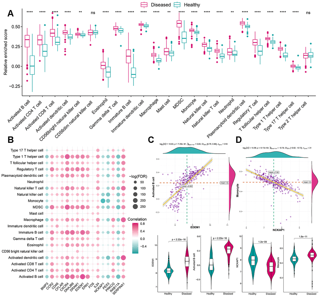
Figure 3.The correlation between infiltrating immunocytes and autophagy genes. (A) The difference in the abundance of each immune microenvironment infiltrating cells between healthy and periodontitis samples. (B) The dot-plot demonstrated the correlations between each dysregulated immune microenvironment infiltration cell type and each dysregulated autophagy genes. (C) The most positive correlated immunocyte-autophagy gene pairs are EDEM1-Activated B cell and the expression status or fraction status are presented by violin-plot at the left panel, indicating a higher expression of EDEM1 and a higher fraction of Activated B cell were found in periodontitis. (D) The most negatively correlated immunocyte-autophagy gene pairs are NCKAP1-Monocyte and the expression status or fraction status are presented by violin-plot at the right panel, indicating there is a lower expression of NCKAP1 in periodontitis and a higher level of the monocyte population.