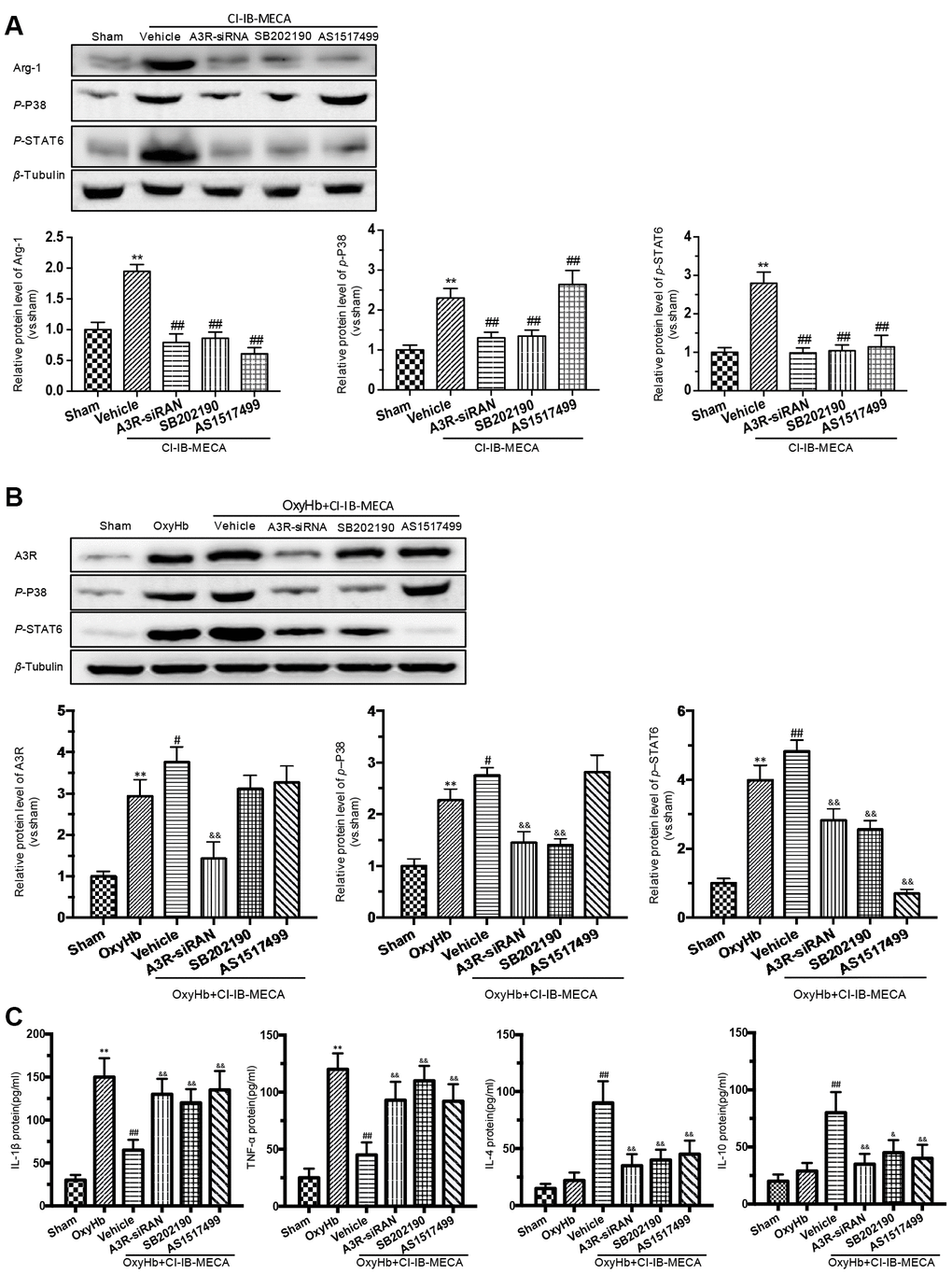
Figure 6.The effects of A3R agonist CI-IB-MECA on microglial polarization are modulated by the A3R/P38/STAT6 pathway. (A) The relationship between the expression of Arg-1 and the A3R/P38/STAT6 axis. To clarify the changes in P38, STAT6 and Arg-1 expression after activation of the adenosine A3 receptor, microglia were treated with CI-IB-MECA alone. Western blots showing the expression of Arg-1 and phosphorylated P38 and STAT6 after pretreatment with A3R siRNA, the P38 blocker (SB202190) and the STAT6 blocker (AS1517499) in microglia treated with CI-IB-MECA. The expression of Arg-1, p-P38 and p-STAT6 under the indicated treatments was also quantified. (B) To further simulate SAH in vitro and demonstrate the role of the A3R/P38/STAT6 pathway, microglia were treated with OxyHb for 24 h in the presence or absence of CI-IB-MECA, A3R siRNA, the P38 blocker (SB202190) or the STAT6 blocker (AS1517499). The protein expression of A3R, p-P38 and p-STAT6 was upregulated after OxyHb stimulation and was further upregulated after the treatment with A3R agonists, which was reversed by A3R siRNA. Inhibition of P38 also decreased STAT6 activation but did not increase A3R expression. (C) The A3R agonist decreased the release of inflammatory cytokines in microglia treated with OxyHb for 24 h. IL-1β and TNF-α increased after treatment with OxyHb and was suppressed by CI-IB-MECA, which was reversed by blocking A3R, P38 or STAT6. IL-4 and IL-10 increased significantly after activation of A3R and decreased after the inhibition of the A3R/P38/STAT6 axis. n=5 in each group. Values are shown as the mean ± SD. **p < 0.01, versus sham group; # p < 0.05, versus OxyHb group; ## p < 0.01, versus CI-IB-MECA group or OxyHb group; & p < 0.05 and && p < 0.01 versus OxyHb+CI-IB-MECA group.