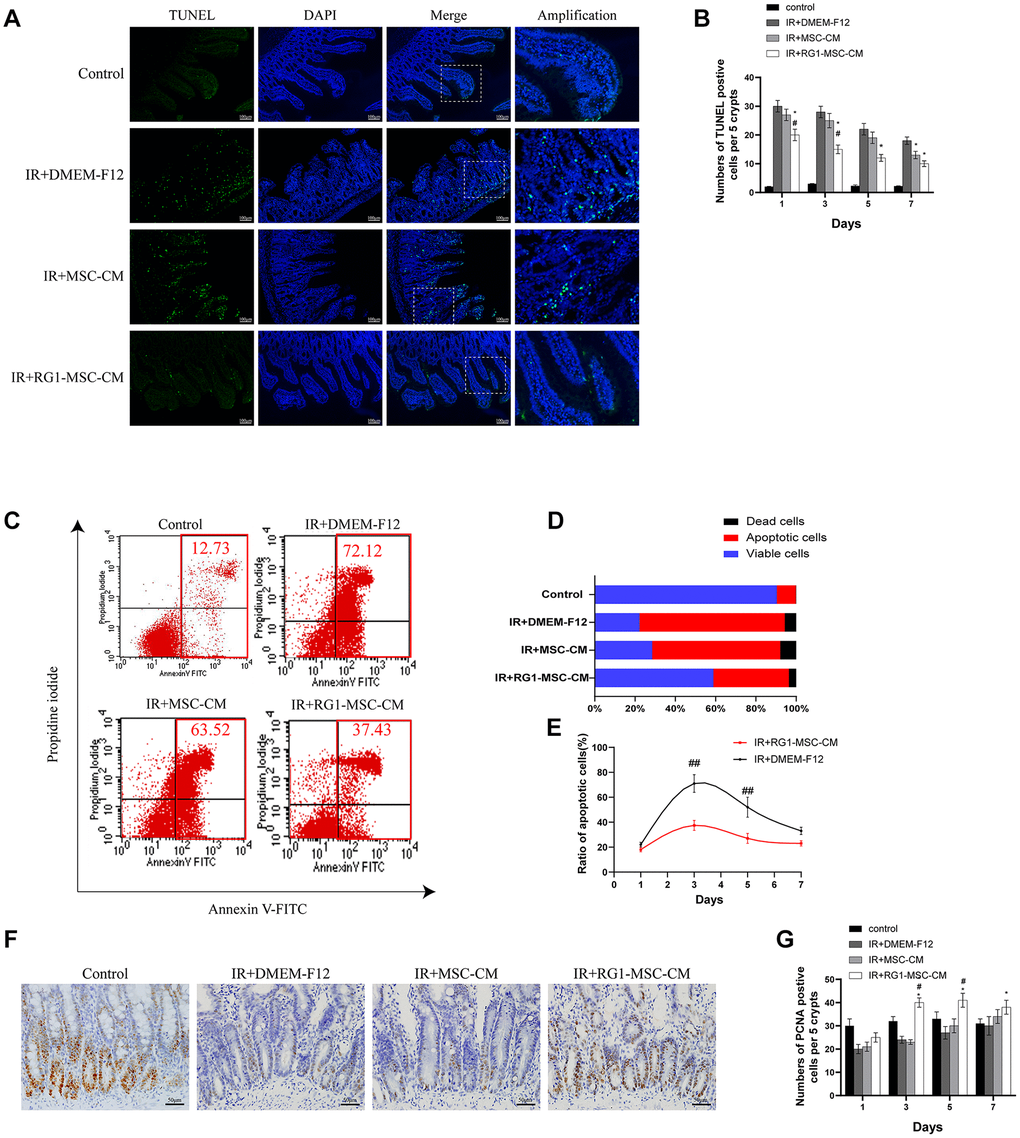
Figure 4.RG1-MSC-CM attenuates apoptosis and promotes proliferation in the intestine. (A) Apoptosis was assayed by TUNEL staining on day 3 of experiment. Scale bars 100 μm. (B) Quantification of TUNEL-positive cells on day 1, 3, 5, 7. n = 3 in each group. The number of positive cells in 5 crypts was scored in 100 crypts per section and reported as mean ± SD. *, P < 0.05 versus IR + DMEM-F12. #, P < 0.05 versus IR + MSC-CM. (C) Apoptosis of IEC-6 was evaluated by flow cytometry after PI/Annexin V staining on day 3 after radiation. The left upper quadrant contains necrotic cells (%); The upper right quadrant contains late apoptotic cells (%); The lower left quadrant contains live cells (%); and the lower right quadrant contains early apoptotic cells (%). (D) The percentage of total apoptotic cells, viable cells and dead cells under each condition are shown. (E) The ratio of apoptotic IEC-6 cells was determined by PI/Annexin V staining at 1, 3, 5, 7 days after radiation. Data represent mean ± SD of three independent experiments. ##, P < 0.05 versus IR+ DMEM-F12. (F) The proliferation of intestinal epithelial cells was examined by immunohistochemical staining with proliferating cell nuclear antigen (PCNA). Intestinal tissue samples were collected and analyzed on day 3 of experiment. Scale bars 50 μm. (G) Quantification of PCNA-positive cells on day 1, 3, 5, 7. n = 3~5 in each group. The number of positive cells in 5 crypts was scored in 100 crypts per section and reported as mean ± SD. *, P < 0.05 versus IR + DMEM-F12. #, P < 0.05 versus IR + MSC-CM. All data were analyzed by t-test or one-way ANOVA except as otherwise indicated.