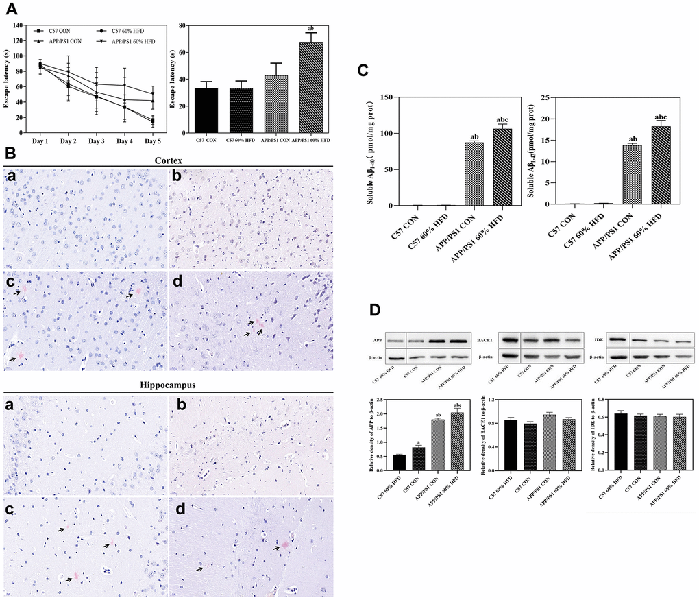
Figure 3.Behavior, Aβ plaque deposit, cortical Aβ content, and APP, BACE1, IDE protein expressions in APP/PS1 and C57 WT mice treated with different diets. APP/PS1 and C57 WT mice were fed with 60% HFD or normal control diets for 6.5 months, then, behavior was test by using MWM (n = 10 for each group); Aβ plaque deposit was measured by using Congo red staining; cortical soluble Aβ1-40 and Aβ1-42 content were test by ELISA method; cortical APP, BACE1 and IDE protein expression was detected by Western blotting (n = 6 at least for each group). Data were expressed as mean ± SE. (A) Escape latency of training and histogram of the time spent in the border area in MWM test. (B) Cortical and hippocampal Aβ plaque deposit. a: control diet-treated C57 WT mice; b: 60% HFD-treated C57 WT mice; c: control diet-treated APP/PS1 mice; d: 60% HFD-treated APP/PS1 mice. Scale bar: 20 μm. (C) Soluble Aβ1-40 and Aβ1-42 content in cortex; a: compared with C57 CON group, P < 0.05; b: compared with C57 60% HFD group, P < 0.05; c: compared with APP/PS1 CON group, P < 0.05. (D) Cortical APP, BACE1 and IDE protein expression. a: compared with C57 60% HFD group, P < 0.05; b: compared with C57 CON group, P < 0.05; c: compared with APP/PS1 CON group, P < 0.05.