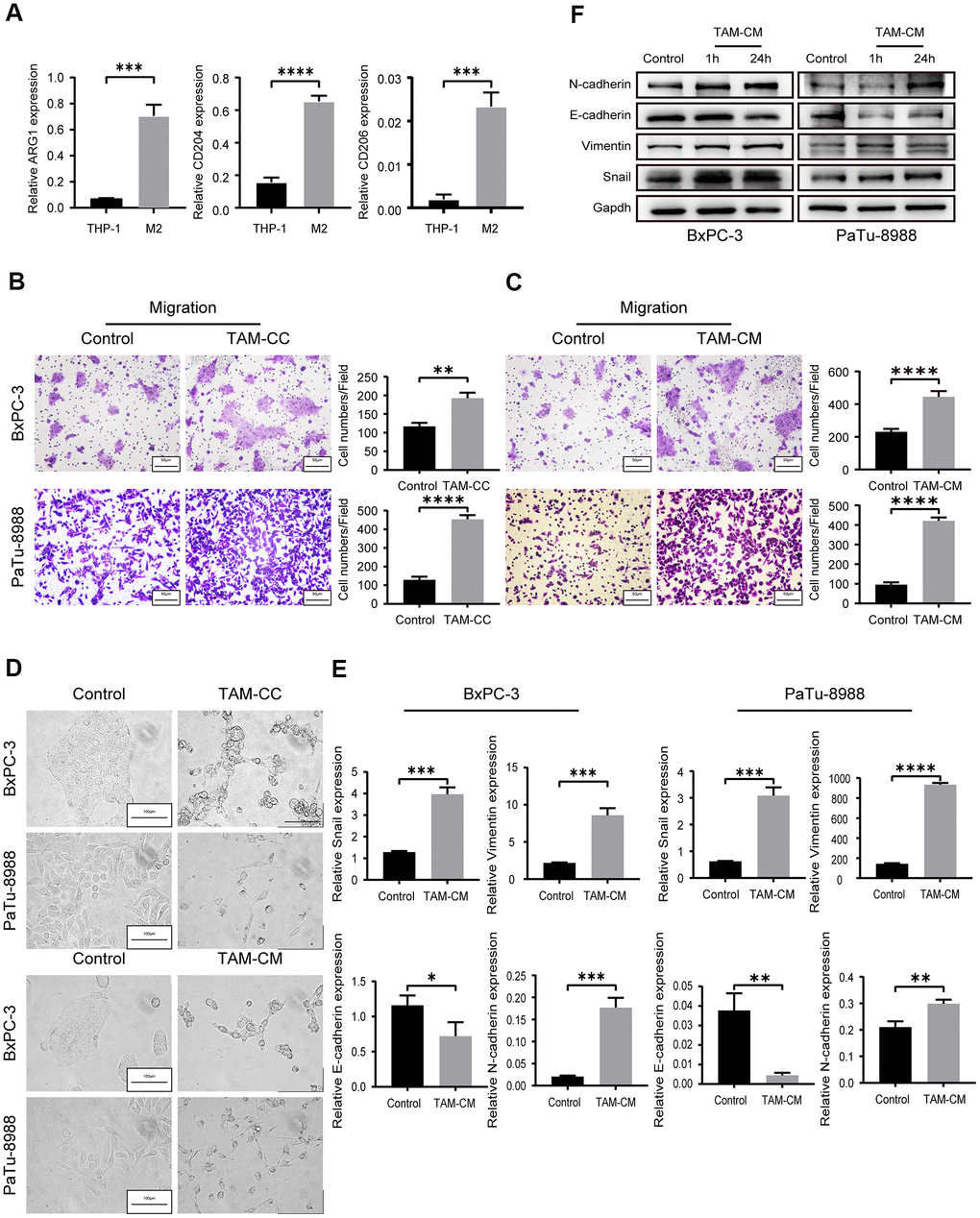
Figure 2.TAMs promote in vitro PDAC cell migration via EMT. (A) QRT-PCR analysis shows relative expression of M2-type macrophage markers such as CD206, CD204, and ARG1 in IL-4-treated THP-1 cells compared to the corresponding controls. ***P < 0.001; ****P < 0.0001. (B) Transwell assay results show the migration and invasiveness of BxPC-3 and PaTu-8988 cells co-cultured with TAMs (TAM-CC) and their corresponding controls. Scale bar = 50 μm; **P < 0.01; ****P < 0.0001. (C) Transwell assay results show the migration and invasiveness of BxPC-3 and PaTu-8988 cells treated with TAM conditional medium (TAM-CM) and their corresponding controls. Scale bar = 50 μm; ****P < 0.0001. (D) Representative images show the morphology of BxPC-3 and PaTu-8988 cells co-cultured with TAMs or TAM-CM and with their corresponding controls. Scale bar = 100 μm. (E) QRT-PCR analysis shows the relative mRNA levels of EMT markers, E-cadherin, N-cadherin, and Vimentin in BxPC-3 and PaTu-8988 cells treated with TAM-CM and their corresponding controls. *P < 0.05; **P < 0.01; ***P < 0.001; ****P < 0.0001. (F) Western blot analyses show the levels of EMT marker proteins, E-cadherin, N-cadherin, and Vimentin in BxPC-3 and PaTu-8988 cells treated with TAM-CM and their corresponding controls.