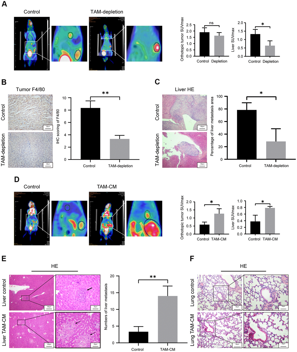
Figure 3.TAMs promote in vivo liver metastasis in the orthotopic PDAC tumor model mice. (A) Positron emission tomography/computed tomography (PET/CT) scanning images show primary pancreatic tumor and liver metastatic lesions in control and TAM-depletion group mice treated with clodronate-liposomes (CLDs). Note: n=6, *P < 0.05. (B) IHC staining analysis shows expression of F4/80 in the primary PDAC tumor tissues from the control and TAM-depletion group mice. Scale bar = 20 μm; **P < 0.01. (C) Representative images show H&E staining analyses of metastatic lesions in the liver sections from control and TAM-depletion group mice treated with clodronate-liposomes (CLDs). Scale bar = 100 μm, *P < 0.05. (D) PET/CT scanning images show primary PDAC tumor and liver metastatic lesions in the control and TAM-CM treatment group mice. Note: n = 6; *P < 0.05. (E) Representative images show H&E staining analyses of metastatic lesions in the liver tissues from control and TAM-CM treatment group mice. Scale bar = 20 μm; **P < 0.01). (F) Representative images show H&E staining analyses of lung tissues from the control and TAM-CM group mice. Scale bar = 50 μm.