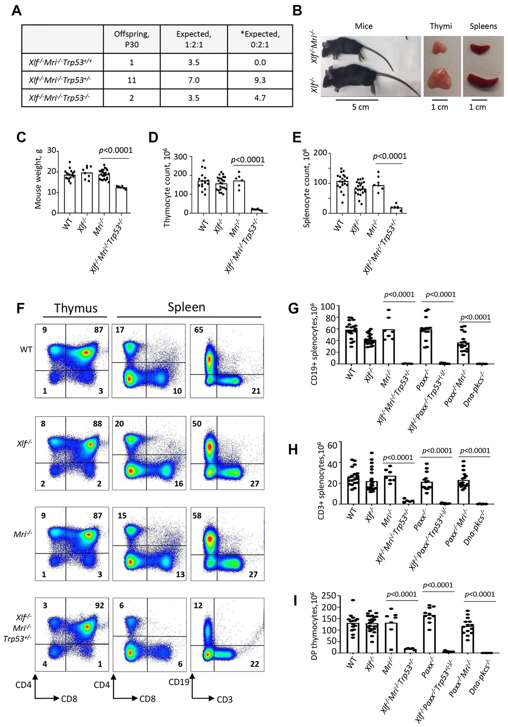
Figure 1.Development of B and T lymphocytes in Xlf-/-Mri-/-Trp53+/- mice. (A) Number of thirty-day-old mice (P30) of indicated genotypes. *Expected distribution assuming lethality. (B) Comparison of body size, thymi and spleens of XLF/MRI-deficient and XLF-deficient mice of the same age. (C) Weights of WT, Xlf-/-, Mri-/-, Xlf-/-Mri-/-Trp53+/- mice. (D, E) Number (×106) of thymocytes (D) and splenocytes (E) in WT, Xlf-/-, Mri-/-, Xlf-/-Mri-/-Trp53+/- mice. (F) Flow cytometric analysis of thymic and splenic T cell subsets and splenic B cells. (G, H, I) Number (×106) of splenic CD19+ B cells (G), splenic CD3+ T cells (H) and thymic CD4+CD8+ double positive (DP) T cells (I) in WT, Xlf-/-, Mri-/-, Xlf-/-Mri-/-Trp53+/-, Paxx-/-, Xlf-/-Paxx-/-Trp53+(-)/- and Paxx-/-Mri-/- mice. Dna-pkcs-/- mice were used as an immunodeficient control. Comparisons between every two groups were made using one-way ANOVA, GraphPad Prism 8.0.1. Xlf-/-Paxx-/-Trp53+(-)/- is a combination of Xlf-/-Paxx-/-Trp53+/- and Xlf-/-Paxx-/-Trp53-/-. Not shown in the graph for (G): WT vs Paxx-/-Mri-/-, p<0.0001 (****), Paxx-/- vs Paxx-/-Mri-/-, p<0.0001 (****), Mri-/- vs Paxx-/-Mri-/-, p<0.0025 (**), Xlf-/- vs Paxx-/-Mri-/-, p=0.9270 (n.s), Xlf-/-Mri-/-Trp53+/- vs Paxx-/-Mri-/-, p<0.0001 (****), and Xlf-/-Paxx-/-Trp53+(-)/- vs Paxx-/-Mri-/-, p<0.0001 (****).