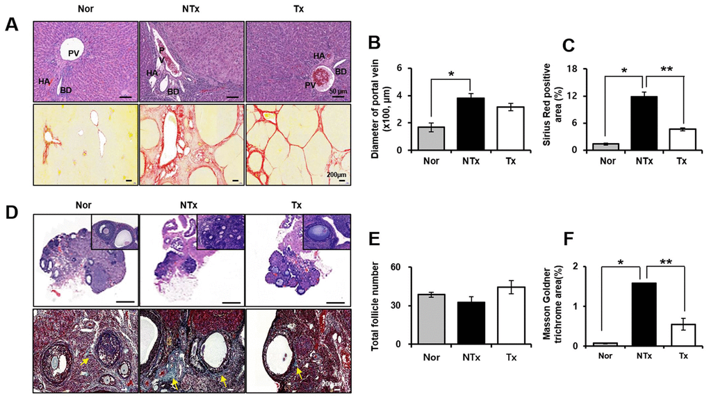
Figure 1.Histological analysis in TAA-injured rat model. H&E (A, upper) and Sirius red (A, lower) was stained and quantification (B, C) from liver in TAA-injured rat model (20x magnification). H&E (D, upper) and Masson Goldner trichrome (D, lower) was stained and quantification (E, F) from ovary in TAA-injured rat model (x20 magnification). Data represent the mean ± S.D. * Significantly different versus Normal (*p<0.05). ** Significantly different versus NTx (**p<0.05). PV, portal vein; HA, hepatic artery; BD, bile duct; SF, secondary follicle; AF, antral follicle; O, oocyte; ATF, atretic follicle.