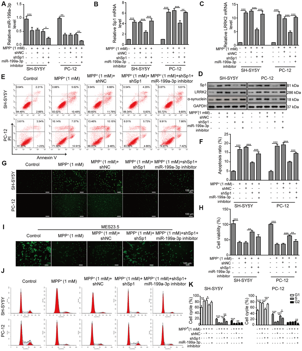
Figure 6.Neuroprotective effects of shSp1 are suppressed by miR-199a-3p inhibition. SH-SY5Y and PC-12 cells were treated with 1 mM MPP+ after transfection with shSp1 or the miR-199a-3p inhibitor. qPCR was performed to assess the expression of (A) miR-199a-3p, (B) Sp1 mRNA, and (C) LRRK2 mRNA. (D) Western blot analysis was conducted to measure the protein expression of Sp1, LRRK2 and α-synuclein under the indicated conditions. (E) Flow cytometry analysis of apoptosis was performed after the cells were transfected with shSp1 and/or the miR-199a-3p inhibitor. (F) Comparison of apoptotic cells in the indicated groups. (G) TUNEL staining was performed to assess cell apoptosis. (H) Cell viability was determined by the CCK-8 assay after the cells were transfected with shSp1 alone or co-transfected with the miR-199a-3p inhibitor. (I) MES23.5 cells were transfected with shNC, shSp1 or the miR-199a-3p inhibitor and treated with 1 mM MPP+. The localization of TUBB3 in MES23.5 cells is shown in the representative images. (J) Cell cycle phases were determined by propidium iodide staining and flow cytometry. (K) Comparison of the cell cycle for the different groups. The data are representative of three experiments. *p <0.05, **p <0.01 and ***p <0.001.