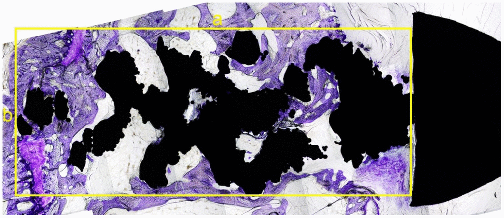
Figure 5.The sagittal slice of histological section of the screw. The black area was the screw (the nut is on the right) and the dyed area (purple) was the bone in-growth tissue. A rectangle was made that can contain part of the screw, without the nut. The longest diameter was measured as a (mm) with the widest diameter as b (mm) (yellow lines). The total area was as S0 = a × b (mm2). The software Image-Pro Plus was used to calculate the tissue area, which was colored by toluidine blue in this rectangle. The percentage of bone in-growth area was defined as S % =S1/S0 x 100%.