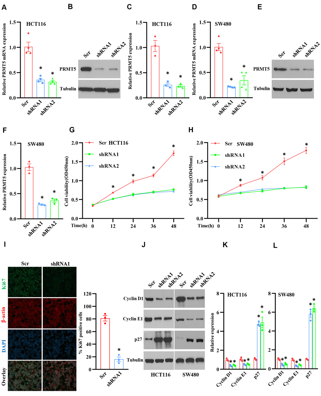
Figure 2.Silencing PRMT5 prevents cell proliferation and cell cycle progression. HCT116 and SW480 cells are transduced by lentivirus containing Scramble and PRMT5-shRNAs. (A) qRT-PCR analysis of PRMT5 mRNA expression level in HCT116 cells. *P < 0.05 vs. Scramble (Scr). (B) PRMT5 protein expression level is detected by Western blotting in HCT116 cells. Representative data is shown. (C) PRMT5 protein expression level is quantified in HCT116 cells. *P < 0.05 vs. Scr. (D) qRT-PCR analysis of PRMT5 mRNA expression level in SW480 cells. * P < 0.05 vs. Scr. (E) PRMT5 protein expression level is analyzed by Western blotting in SW480 cells. (F) PRMT5 protein expression level is quantified in SW480 cells. *P < 0.05 vs. Scr. (G, H) Cell viability is measured during different time points in HCT116 and SW480 cells (n=4). *P < 0.05 vs. Scr. (I) Immunostaining of ki67, a cell proliferation marker, in HCT116 cells. Representative pictures were shown. Scale bar = 50μm. The Ki67 positive cells were quantified in the indicated groups. For each group, 1000 cells were counted. n=3, *P < 0.05 vs. Scr. (J) Western blot analysis of cyclin D1, cyclin E1, and p27 protein expression level in HCT116 and SW480 cells. Representative data is shown. (K, L) Indicated proteins are quantified in HCT116 and SW480 cells. *P < 0.05 vs. Scr. Red bar = Scr, Blue bar = shRNA1, and Green bar = shRNA2.