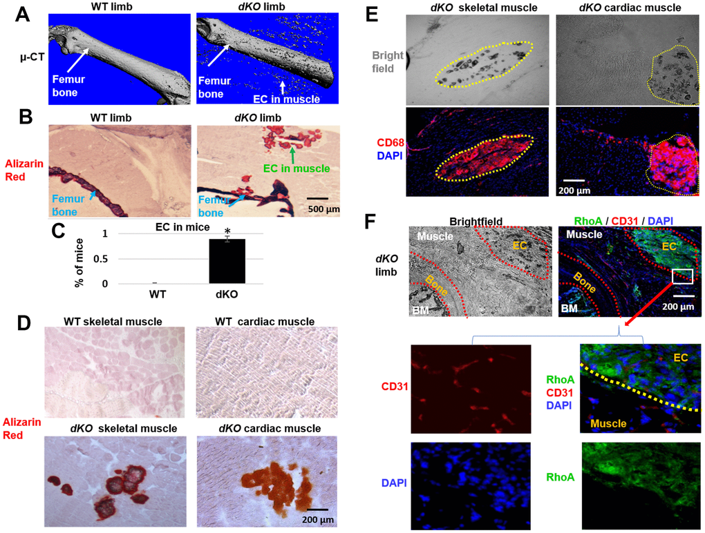
Figure 1.Increased macrophage accumulation at sites of ectopic calcification in skeletal muscle of dKO mice. (A). Micro-CT scanning results indicating increased ectopic calcification and osteoporosis in the hindlimb of dKO mice, compared to WT mice. (B) Alizarin Red staining of hindlimb tissue sections validate the presence of ectopic calcification in dKO mice. (C) Percent of mice that exhibit ectopic calcification (EC) in hindlimbs of dKO mice and WT mice. (D) Alizarin Red staining of skeletal muscle (gastrocnemius) and cardiac muscle (septum) showing ectopic calcification (EC). (E) Immunostaining of dKO skeletal muscle and heart sections with CD68 antibody and bright field imaging showing the extensive accumulation of CD68+ macrophages at the sites of ectopic calcification (EC). (F) Immunostaining of dKO hindlimb sections with RhoA and CD31 antibodies and bright field imaging showing the increased accumulation of RhoA+ cells at the sites of ectopic calcification (EC). n=8 for both WT and dKO mice (8-week old).*=p<0.05.