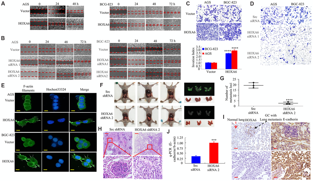
Figure 4.In vitro and in vivo, the effects of HOXA6 on migration and invasion in GC cells. (A, B) Wound healing assay in GC cells transfected with HOXA6 plasmid or HOXA6 siRNA. Relative wound closure was calculated in experiments performed in triplicate. (C, D) Transwell assay conducted in GC cells transfected with HOXA6 plasmid or HOXA6 siRNA. The number of invading cells is shown. Cells in 5 independent symmetrical visual fields from three independent experiments were counted under a microscope. ****, P < 0.001. (E) Stable HOXA6 transfectants were stained with rhodamine-phallotoxin to reveal F-actin filaments by fluorescence microscopy. (F) Whole-body fluorescence imaging showing GC progression in mice (n = 3). Metastatic loci in the lungs are detected in the image. (G) Metastatic loci in the lung were calculated. ****, P < 0.001. (H) Lung sections were stained with haematoxylin and eosin. (I) The expression of HOXA6 and E-cadherin in the lung metastasis in GC was determined by IHC. (J) qRT-PCR was detected the expression of E-cadherin in lung tumours derived from AGS cells. ***, P < 0.01. Scale bars, 50 μm in E, 100 μm in H and I.