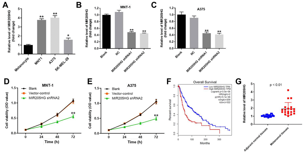
Figure 1.Knockdown of MIR205HG significantly decreased the viability of melanoma cells. (A) QRT-PCR analysis shows the expression of MIR205HG in melanocytes, A375, MNT-1, and SK-MEL-28 cells. (B, C) QRT-PCR analysis shows the expression of MIR205HG in (B) MNT-1 and (C) A375 cells transfected with MIR205HG shRNA1, shRNA2 or sh-NC for 24 h. (D, E) CCK-8 assay analysis results show the viability of blank, vector-control, and MIR205HG shRNA2-transfected MNT-1 and A375 cells for 0, 24, 48 or 72 h. (F) TCGA database analysis shows the correlation between MIR205HG expression and survival rates of melanoma patients. The analysis included the data from 531 patients with melanoma. Among the patients with melanoma, 209 patients had high expression of MIR205HG, while the others had low level of MIR205HG. (G) QRT-PCR analysis shows the expression of MIR205HG in paired melanoma and adjacent normal skin tissues (n=30). Note: All experiments were performed at least thrice independently. *P<0.05 and **P<0.01 vs. control.