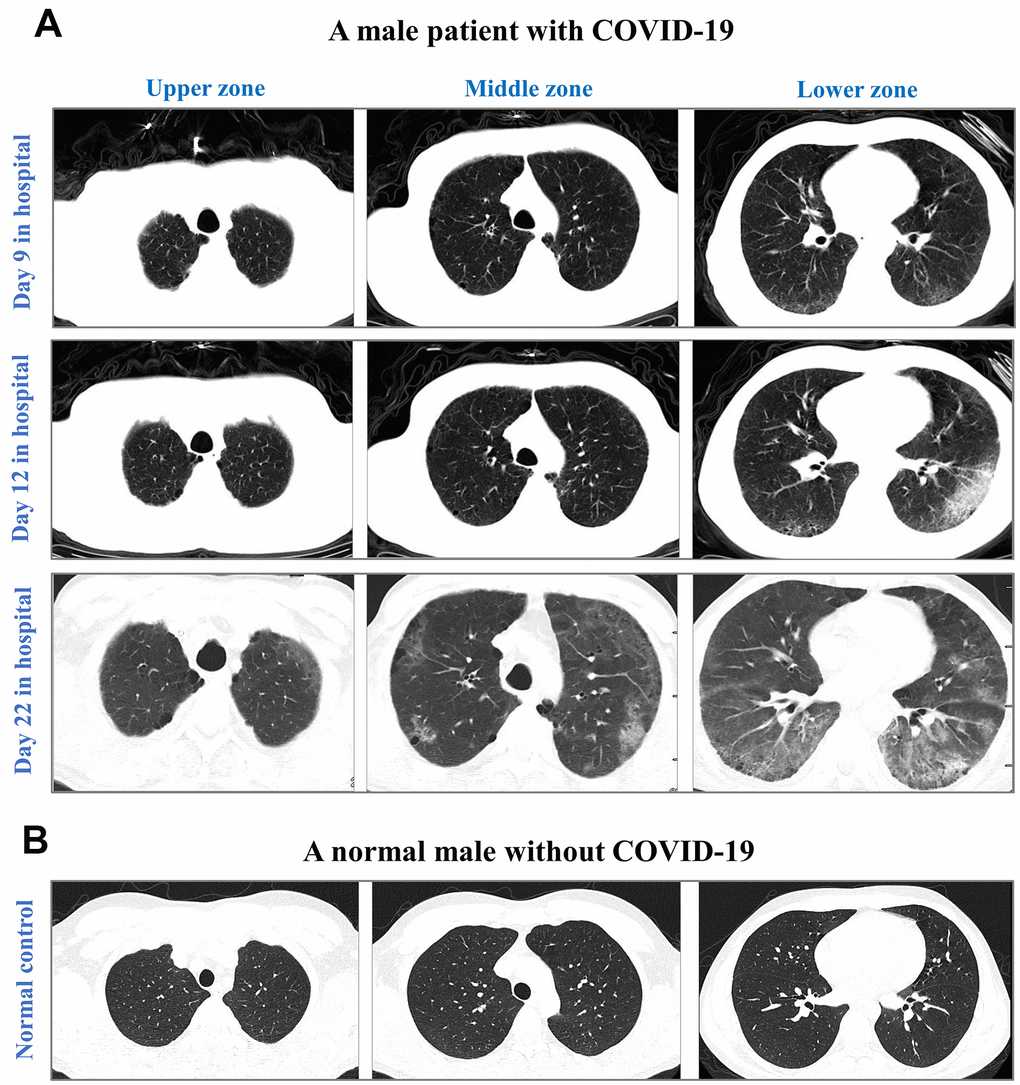
Figure 4.CT images from an elderly male with laboratory-confirmed COVID-19 (A) and a normal male without COVID-19 (B). CT images were illustrated in the upper zone (above the carina), the middle zone (below the carina up to the inferior pulmonary vein), and the lower zone (below the inferior pulmonary vein). After 9-day hospitalization, CT images showed minor ground-glass opacity (GGO) in subpleural areas of the lower left and right lobes in the COVID-19 male. After 12-day hospitalization, CT images showed progressing GGOs and newly-appeared reticulation. The vascular enlargement was observed in the lesion of the lower left lung. A small amount of bilateral pleural effusion was newly developed. After 22-day hospitalization, CT images showed progressing lesion with multiple newly-appeared GGO in both lungs, predominantly located in subpleural areas of lower lobes. Bronchiectasis of the anterior internal basal segment of the left lower lung was visible. Progressing bilateral pleural effusion was identified. The patient passed away after 24 days of hospitalization in The First Hospital of Changsha.