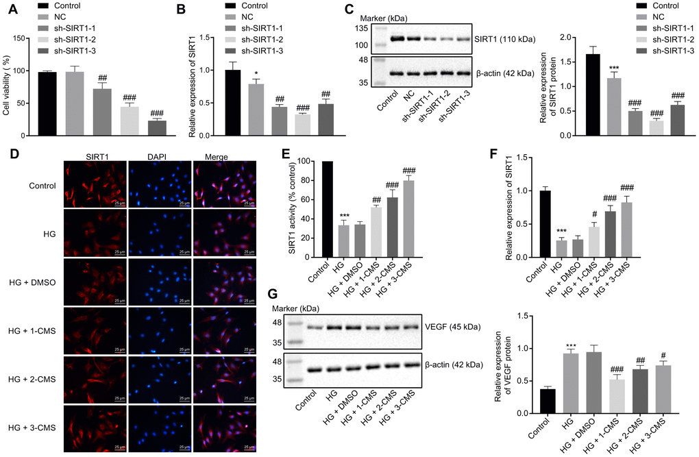
Figure 4.CMS activated the expression of SIRT1 in HG-treated hRMECs. hRMECs were transfected with sh-SIRT1-1, sh-SIRT1-2, or sh-SIRT1-3, and HG-exposed hRMECs were treated with DMSO, 1-CMS, 2-CMS, or 3-CMS, respectively. (A) Cell viability assessed by CCK-8 assay. (B) SIRT1 expression pattern determined by RT-qPCR in hRMECs, normalized to β-actin. (C) Representative Western blots of SIRT1 protein and its quantitation in hRMECs, normalized to β-actin. (D, E) Representative images (× 400) (scale bar = 25 μm) (D) as well as SIRT1 activity and nuclear accumulation in hRMECs (E) detected by immunofluorescence staining. (F) Expression pattern of SIRT1 as determined by RT-qPCR in hRMECs, normalized to β-actin. (G) Representative Western blots of VEGF protein and its quantitation in hRMECs, normalized to β-actin. *p < 0.05, **p < 0.01, ***p < 0.001, compared to the control cells, and #p < 0.05, ##p < 0.01, ###p < 0.001, compared to cells stimulated with NC or HG + DMSO. The results were measurement data and expressed as mean ± standard deviation. Comparisons between multiple groups were analyzed by one-way ANOVA with Tukey’s post hoc test. The cell experiments were repeated three times independently. NC, negative control; CMS, coumestrol, HG, high glucose; DMSO, dimethyl sulfoxide; SIRT1, sirtuin 1; RT-qPCR, reverse transcription-quantitative polymerase chain reaction; VEGF, vascular endothelial growth factor; ANOVA, analysis of variance.