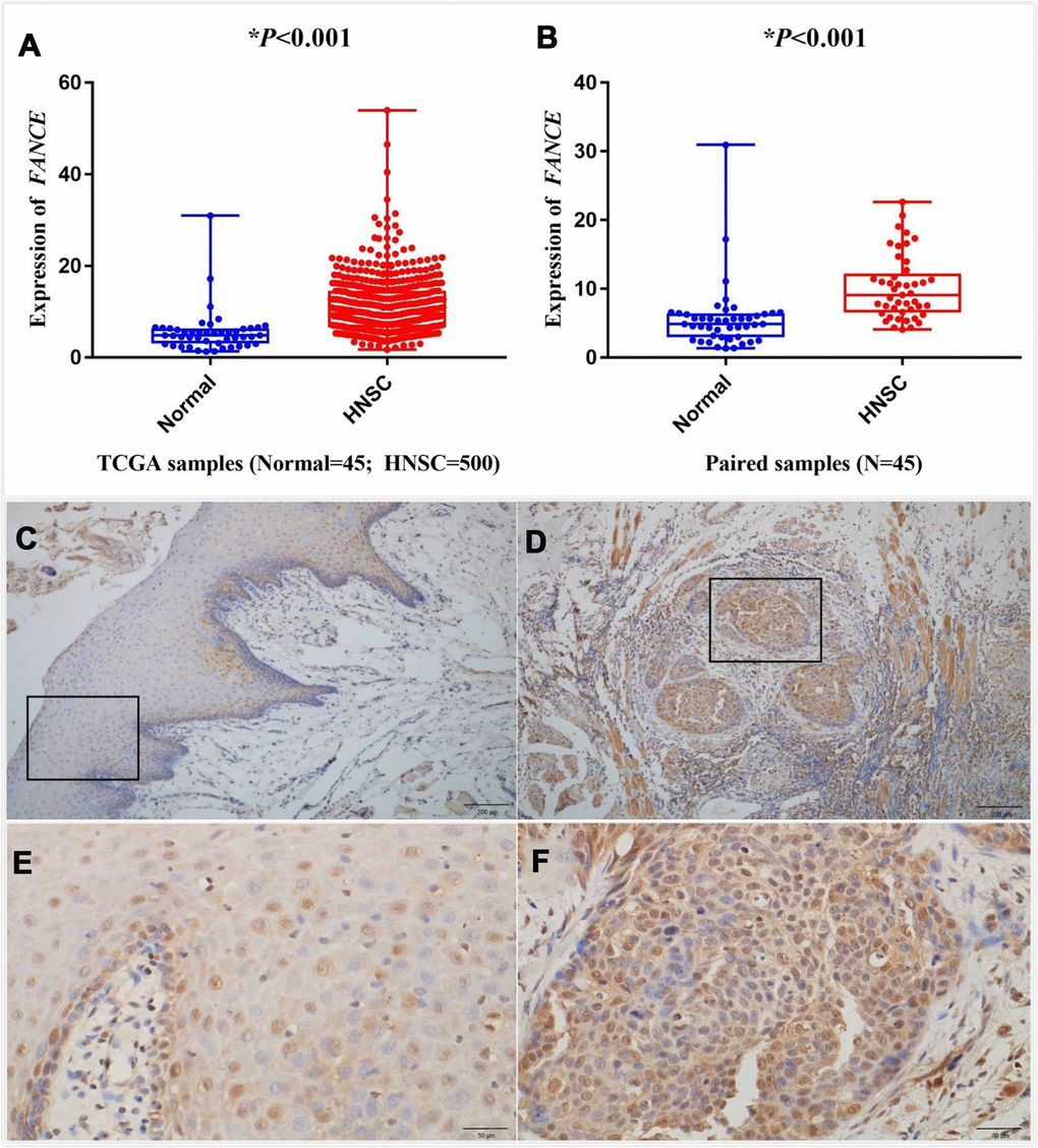
Figure 6.Analysis of differential expression of FANCE in HNSC. Representative IHC staining (200x) performed for detecting the expression of FANCE from adjacent normal tissues and tumor-tissue specimens of HNSC patients from human protein atlas (https://www.proteinatlas.org/). (A) FANCE expression was compared between HNSC tissues and normal tissues based on the TCGA database. FANCE expression in HNSC (n = 500) was significantly increased than that in normal patients (n = 45). (B) The level of FANCE expression was compared between the paired samples. Matching cancer tissues and adjacent normal tissues of the same patient also showed that FANCE expression was significantly up-regulated in cancer tissues. (C–F) C and E showed normal tissue, while D and F were HNSC tissue. Immunohistochemical staining was performed to detecting the expression of FANCE in adjacent normal tissues (C, x200 and E, x400) and tumor tissue specimens (D, x200 and F, x400) of patients with HNSC.
Figure 6 — Comprehensive analysis of macrophage-related multigene signature in the tumor microenvironment of head and neck squamous cancer | Aging