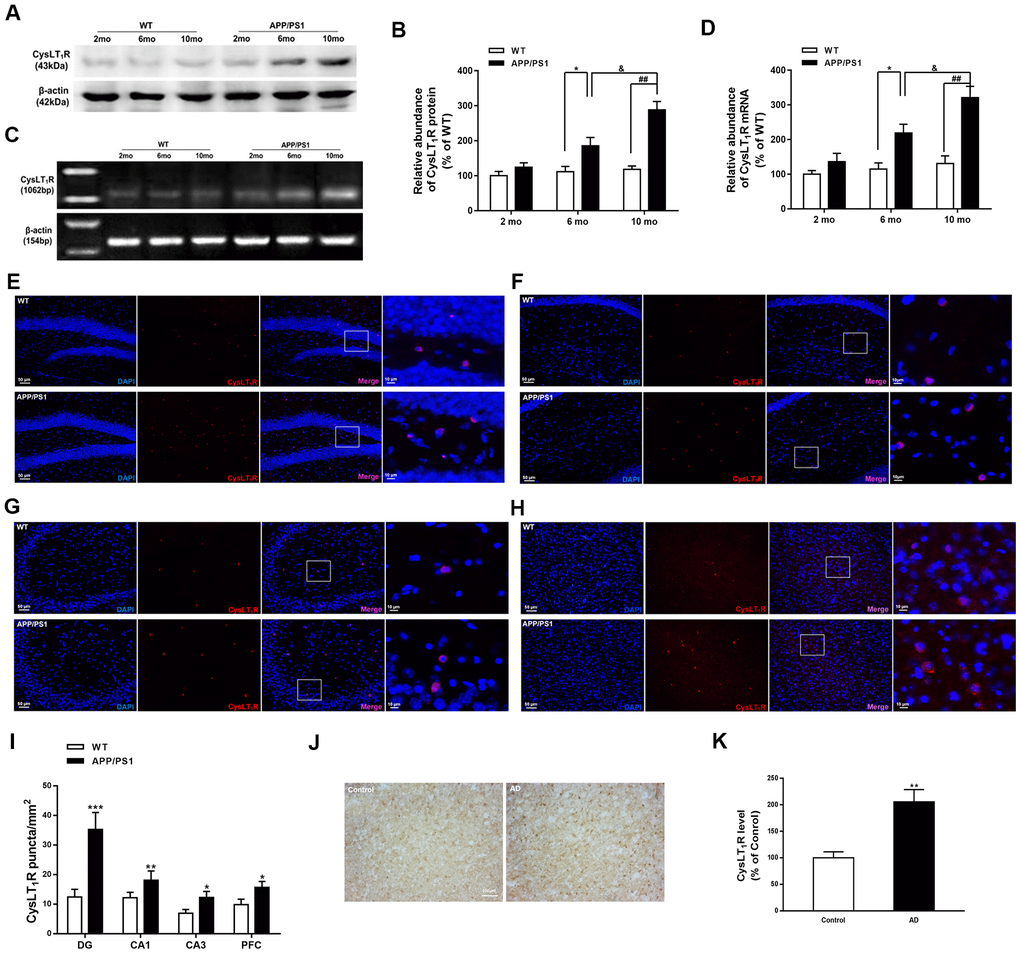
Figure 1.CysLT1R expression is upregulated in APP/PS1 mice and AD patients. (A) Representative immunoblots of CysLT1R protein in the hippocampus of APP/PS1 mice and WT mice at the age of 2, 6 and 10 months. (B) Quantification of CysLT1R protein levels was expressed as the ratio (in %) of WT group. Data are expressed as mean ± SEM, n = 4, *P < 0.05 vs. 6-month-old WT mice; ##P < 0.01 vs. 10-month-old WT mice; &P < 0.05 vs. 6-month-old APP/PS1 mice. (C) RT-PCR detection of CysLT1R mRNA in the hippocampus of APP/PS1 mice and WT mice at the age of 2, 6 and 10 months. (D) Quantification of CysLT1R mRNA levels was expressed as the ratio (in %) of WT group. Data are expressed as mean ± SEM, n = 4, *P < 0.05 vs. 6-month-old WT mice; ##P < 0.01 vs. 10-month-old WT mice; &P < 0.05 vs. 6-month-old APP/PS1 mice. Immunofluorescence images of CysLT1R expression in the hippocampal DG (E), CA1 (F), CA3 (G), and prefrontal cortex (H) in APP/PS1 mice and WT mice. Scale bar = 50 μm. (I) Quantification of CysLT1R in the brain sections of mice. Data are expressed as mean ± SEM, n = 4, *P < 0.05, **P < 0.01, ***P < 0.01 vs. WT mice. (J) CysLT1R levels in the brain sections from post-mortem AD patients and normal controls by immunohistochemical analyses. Scale bar = 100 μm. (K) Quantification of CysLT1R in the sections of human postmortem brains. Data are expressed as mean ± SEM, n = 4, **P<0.01 vs. control.