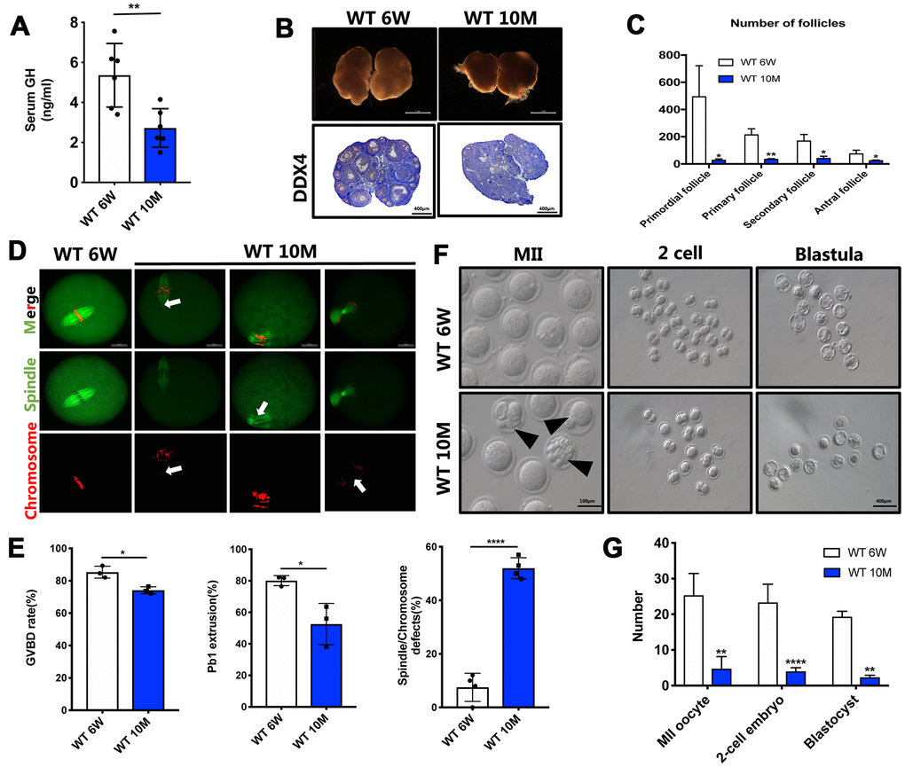
Figure 1.The GH levels and oocyte quality were declined in aged mice. (A) The GH levels in the peripheral blood were measured in young (n = 6) and aged (n = 6) mice. (B) Micrographs of young and aged WT mouse ovaries (Scale bar, 1 mm) and HE staining of these ovaries (Scale bar, 100μm, 500 μm). (C) Follicle counts in 6-week (n = 3) and 10-month (n = 3) old WT mice. (D) Chromosomes misalignment and spindle defects (arrowheads) in aged oocytes. The oocytes were stained with α-tubulin (green) and propidium iodide (PI) (red) respectively. Scale bar, 50 μm. (E) Left: The rate of GVBD and Pb1 extrusion were recorded after 4 h and 14 h of culture in M2 medium respectively. Right: Percentages of oocytes with spindle defects in young (n = 97) and aged mice (n = 104). (F) Representative images of MII oocytes collected from young (n = 76) and aged (n = 19) mice and IVF outcomes from these two groups. Black arrowheads point to abnormal oocytes. Scale bar, 100 μm, 400 μm. (G) Quantification of MII oocytes, 2-cell embryo and blastocyst from young and aged mice. Data are presented as mean ± SD. *P < 0.05, **P < 0.01, ****P < 0.0005.