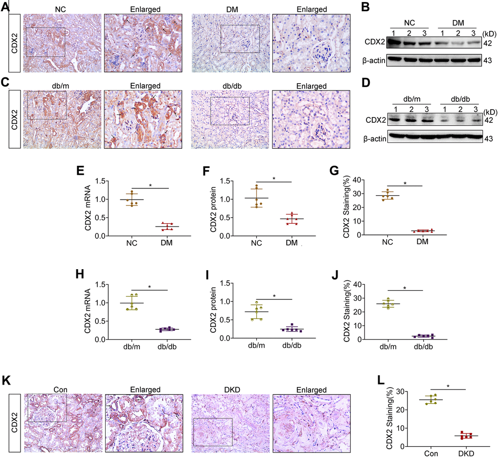
Figure 1.Kidney CDX2 is downregulated in DKD. T1D mice (DM group) and controls were submitted to euthanasia at 16 weeks of age (6 weeks after the establishment of the T1D mouse model), and T2D mice (db/db group) and controls were submitted to euthanasia at 18 weeks of age. (A, B) Immunohistochemical staining (A) and immunoblot (B) for CDX2 detection in T1D mice and controls. (C, D) Immunohistochemical staining (C) and immunoblot (D) for CDX2 detection in T2D mice and controls. (E–G) Immunohistochemical-positive staining density of CDX2 was analyzed in each group from 6 random fields (200×). Quantitation of mRNA amounts (E), Western blot bands (F) and immunohistochemical signals (G) of CDX2 in T1D mice kidney tissues and controls. (H–J) Quantitation of mRNA amounts (H), Western blot bands (I) and immunohistochemical signals (J) of CDX2 in T2D mice kidney tissues and controls. (K, L) Immunohistochemical staining (K) and quantitative analysis (L) of CDX2 in DKD patients kidney tissues and controls. CDX2 is expressed in the cytoplasm and nucleus of renal tubular epithelial cells in the renal cortex (black arrow) (magnification, ×200); enlarged box area (magnification,×400). All data are mean±SD from three independent experiments. n=6; *P<0.05.